. 2013 May 30;497(7451):633-7.
doi: 10.1038/nature12138. Epub 2013 May 12.

Macropinocytosis of protein is an amino acid supply route in Ras-transformed cells

Affiliations

Macropinocytosis of protein is an amino acid supply route in Ras-transformed cells

Cosimo Commisso et al. Nature. .

Abstract

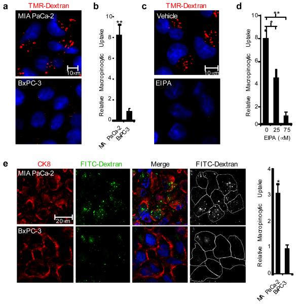
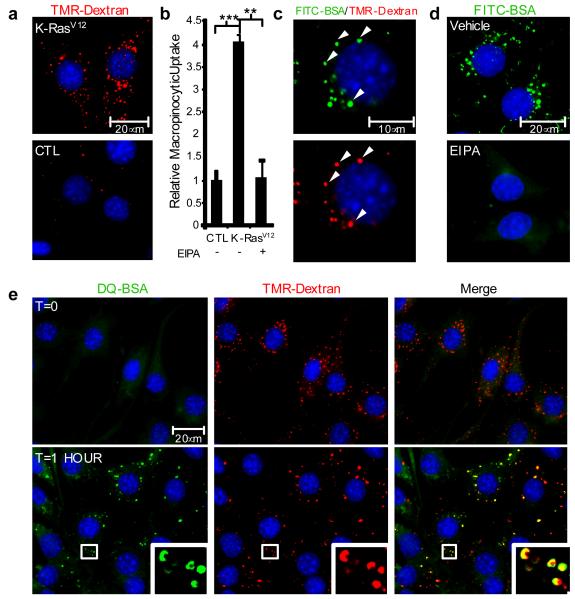
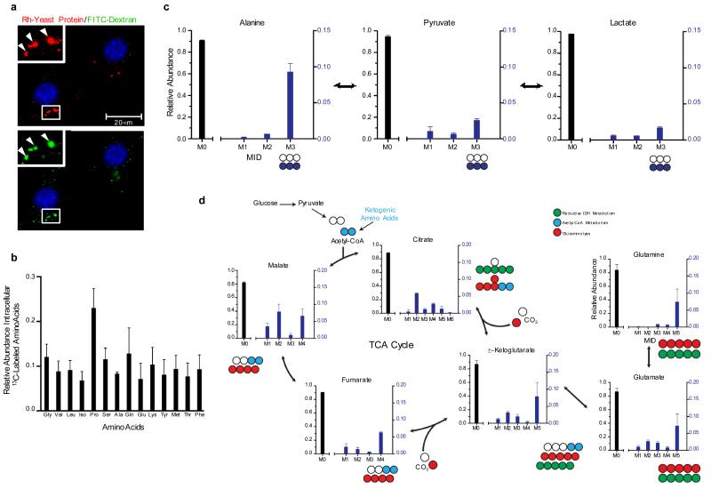
Macropinocytosis is a highly conserved endocytic process by which extracellular fluid and its contents are internalized into cells through large, heterogeneous vesicles known as macropinosomes. Oncogenic Ras proteins have been shown to stimulate macropinocytosis but the functional contribution of this uptake mechanism to the transformed phenotype remains unknown. Here we show that Ras-transformed cells use macropinocytosis to transport extracellular protein into the cell. The internalized protein undergoes proteolytic degradation, yielding amino acids including glutamine that can enter central carbon metabolism. Accordingly, the dependence of Ras-transformed cells on free extracellular glutamine for growth can be suppressed by the macropinocytic uptake of protein. Consistent with macropinocytosis representing an important route of nutrient uptake in tumours, its pharmacological inhibition compromises the growth of Ras-transformed pancreatic tumour xenografts. These results identify macropinocytosis as a mechanism by which cancer cells support their unique metabolic needs and point to the possible exploitation of this process in the design of anticancer therapies.


Figures

Figure 1
Figure 1. Oncogenic K-Ras-expressing pancreatic cancer cells display elevated levels of macropinocytosis both in culture and in vivo
a, A macropinocytosis uptake assay utilizing TMR-dextran as a marker of macropinosomes (red) indicates that MIA PaCa-2 cells display elevated levels of macropinocytosis compared to BxPC-3 cells. DAPI staining (blue) identifies nuclei. b, Data are expressed as arbitrary units and are presented relative to the values obtained for BxPC-3 cells. c, Macropinocytic uptake in MIA PaCa-2 cells treated with either vehicle (DMSO) or 75 μM EIPA. d, Quantification of macropinocytic uptake in MIA PaCa-2 cells treated with 0, 25 or 75 μM EIPA. Data are presented relative to the values obtained for the 75 μM condition. e, Visualization and quantification of macropinocytosis in vivo. Representative images from sections of FITC-dextran (green) injected tumor xenografts stained with anti-CK8 (red). Cell boundaries (white outline) were delineated based on the CK8 staining. Data are presented relative to the values obtained for the BxPC-3 tumors. For all graphs, error bars indicate mean +/− SEM for n=3 independent experiments with at least 300 cells scored per experiment. Statistical significance was determined via t-test; *p<0.05, **p<0.01.
Figure 2
Figure 2. Oncogenic K-Ras-induced macropinocytosis in NIH 3T3 cells mediates the internalization of extracellular albumin that is subsequently targeted for proteolytic degradation
a, TMR-dextran (red) is internalized at higher levels in NIH 3T3 [K-RasV12] cells (K-RasV12, top panel) compared to untransformed control cells (CTL, bottom panel). b, Quantification of macropinocytic uptake in control cells and NIH 3T3 [K-RasV12] cells incubated with vehicle (DMSO) or with 75 μM EIPA. Data are presented relative to the values obtained for the untransformed control cells. Error bars indicate mean +/− SEM for n=3 independent experiments with at least 300 cells scored per experiment. Statistical significance was determined via t-test; **p<0.01, ***p<0.001. c, FITC-BSA (green) is internalized into discrete puncta that co-localize (white arrowheads) with TMR-dextran (red). d, FITC-BSA uptake is abrogated by treatment with 75 μM EIPA. e, Analysis of DQ-BSA fluorescence in NIH 3T3 [K-RasV12] cells that were co-incubated with DQ-BSA (green) and TMR-dextran (red) and fixed either immediately (T=0) or following a 1 hour chase. The fluorescent signal emanating from DQ-BSA (T=1 HOUR) is an indication of albumin degradation. Insets represent a higher magnification of the boxed areas. Images shown in c, d and e are representative of at least 3 independent experiments.
Figure 3
Figure 3. Macropinocytic uptake of extracellular protein drives the accumulation of catabolic intermediates and entry of protein-derived amino acids into central carbon metabolism
a, Rhodamine-labeled (Rh) yeast protein (red) is internalized into puncta (arrowheads) that co-localize with FITC-dextran (green). Insets represent a higher magnification of the boxed areas. b, Uniformly 13C-labeled intracellular amino acid pools were detected in NIH 3T3 [K-RasV12] cells after culture in low glutamine-containing medium (0.2 mM) supplemented with 2% 13C-labeled yeast protein. c, Protein-derived alanine enters central carbon metabolism upon transamination to pyruvate and pyruvate can be directly converted to lactate. M3 reflects fully labeled alanine, pyruvate and lactate, while M0 abundances reflect metabolites with no 13C label. M1 and M2 represent partially labeled species that are not present in significant amounts. d, Atom transition map depicting a model for the entry of amino acid-derived carbons into the TCA cycle and isotopic labeling of various metabolites. Open circles represent unlabeled carbon, and different colored circles highlight labeling patterns that correspond to specific pathways. For all graphs, error bars indicate mean +/− SD for three independent experiments. IDH, isocitrate dehydrogenase; MID, mass isotopomer distribution
Figure 4
Figure 4. Macropinocytosis is required for albumin-dependent cancer cell proliferation in vitro and for tumor growth in vivo
a, The compromised proliferation of oncogenic Ras-expressing cells resulting from growth in media containing subphysiological concentrations of glutamine (0.2 mM, 0.2Q) is reversed by supplementation with 2% albumin (0.2Q+Alb) and this effect is inhibited by treatment with 25 μM EIPA (0.2Q+Alb+EIPA). Total viable cell counts were measured via MTT assay after 6 days of growth. Data are presented relative to the values obtained for the 0.2Q condition. b, The effects of EIPA treatment (25 μM) are suppressed by increasing the glutamine levels in the growth media to the indicated concentrations (i.e. 0.5Q indicates 0.5 mM glutamine) or the addition to the medium of 7 mM dimethyl α-ketoglutarate (KG). Data are presented relative to the values obtained for the 0.2Q+Alb condition. For both (a) and (b), error bars indicate mean +/− SEM for n=3 independent experiments. Statistical significance was determined via t-test; *p<0.05, **p<0.01. c-e, EIPA inhibits macropinocytosis in vivo and reduces tumor growth in a subcutaneous heterotopic xenograft model of pancreatic cancer. c, Representative images from sections of FITC-dextran (green) injected MIA PaCa-2 tumor xenografts from mice treated with EIPA or vehicle only controls after 7 days of treatment. The human pancreatic cancer cells are marked by anti-CK8 staining (red). d, Representative digital photographs of dissected tumors from mice treated with EIPA or vehicle only controls. e, Waterfall plots indicating the percent change in tumor volume after seven days of treatment relative to baseline (Day 0 of treatment) for tumors derived from MIA PaCa-2 cells or BxPC-3 cells.

Comment in

  • Metabolism: Taking it all in.
    Seton-Rogers S. Seton-Rogers S. Nat Rev Cancer. 2013 Jul;13(7):438. doi: 10.1038/nrc3550. Epub 2013 Jun 6. Nat Rev Cancer. 2013. PMID: 23739049 No abstract available.

References

    1. Bar-Sagi D, Feramisco JR. Induction of membrane ruffling and fluid-phase pinocytosis in quiescent fibroblasts by ras proteins. Science. 1986;233:1061–1068. - PubMed
    1. Porat-Shliom N, Kloog Y, Donaldson JG. A unique platform for H-Ras signaling involving clathrin-independent endocytosis. Mol Biol Cell. 2008;19:765–775. - PMC - PubMed
    1. Walsh AB, Bar-Sagi D. Differential activation of the Rac pathway by Ha-Ras and K-Ras. J Biol Chem. 2001;276:15609–15615. - PubMed
    1. Lopez-Crapez E, Chypre C, Saavedra J, Marchand J, Grenier J. Rapid and large-scale method to detect K-ras gene mutations in tumor samples. Clinical chemistry. 1997;43:936–942. - PubMed
    1. Aoki K, Yoshida T, Sugimura T, Terada M. Liposome-mediated in vivo gene transfer of antisense K-ras construct inhibits pancreatic tumor dissemination in the murine peritoneal cavity. Cancer Res. 1995;55:3810–3816. - PubMed

Publication types

MeSH terms