Review
. 2013 Jun;12(6):465-83.
doi: 10.1038/nrd4023. Epub 2013 May 13.

Pharmacological approaches to restore mitochondrial function

Affiliations
Review

Pharmacological approaches to restore mitochondrial function

Pénélope A Andreux et al. Nat Rev Drug Discov. 2013 Jun.

Abstract

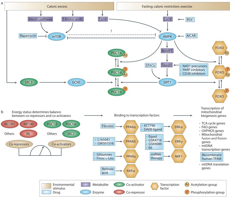
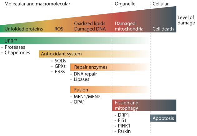
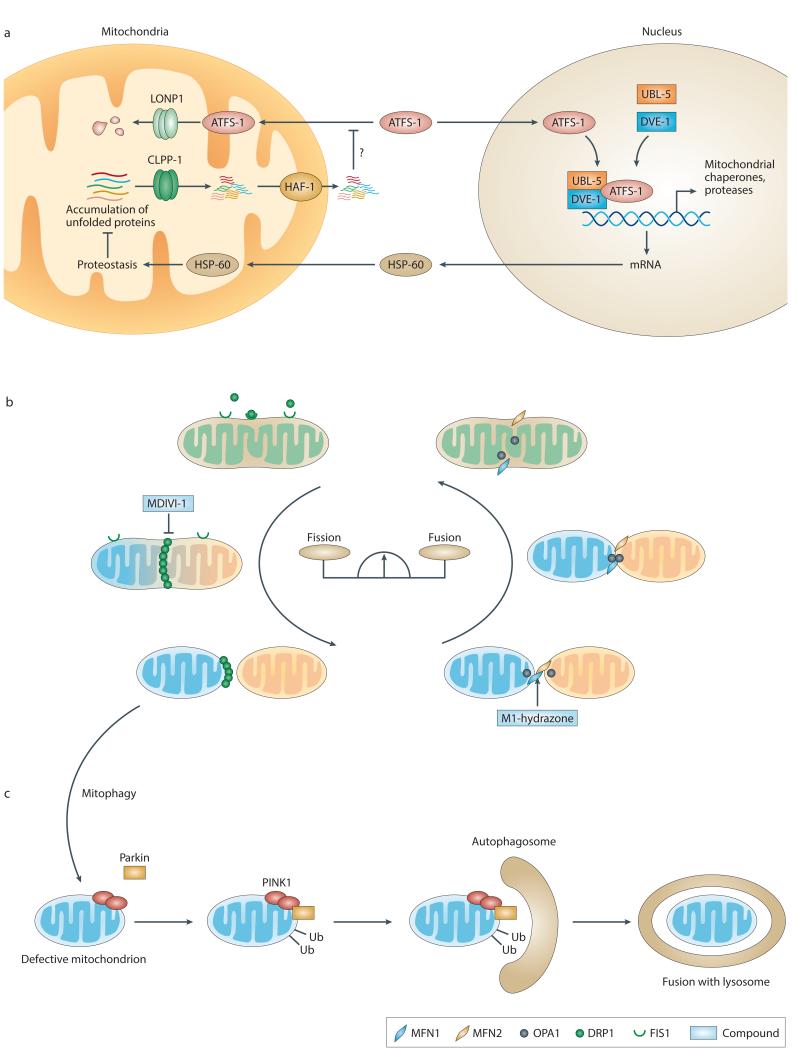
Mitochondrial dysfunction is not only a hallmark of rare inherited mitochondrial disorders but also implicated in age-related diseases, including those that affect the metabolic and nervous system, such as type 2 diabetes and Parkinson's disease. Numerous pathways maintain and/or restore proper mitochondrial function, including mitochondrial biogenesis, mitochondrial dynamics, mitophagy and the mitochondrial unfolded protein response. New and powerful phenotypic assays in cell-based models as well as multicellular organisms have been developed to explore these different aspects of mitochondrial function. Modulating mitochondrial function has therefore emerged as an attractive therapeutic strategy for several diseases, which has spurred active drug discovery efforts in this area.


Figures

Figure 1
Figure 1. Pharmacological approaches targeting mitochondrial biogenesis
Pharmacological approaches for targeting mitochondrial biogenesis. a | Upstream sensors of energy status. Energy stress promotes the activation of AMP-activated protein kinase (AMPK) via an increase in the AMP/ATP ratio, whereas energy excess activates the mammalian target of rapamycin (mTOR) pathway either via an increase in levels of amino acids or via the activation of insulin signalling. Rapamycin, 5-aminoimidazole-4-carboxamide riboside (AICAR) and resveratrol (RSV) simulate energy crisis by inhibiting mTOR or activating AMPK. AMPK-mediated phosphorylation of co-factors (such as PPARγ co-activator 1α (PGC1α)) and transcription factors (such as forkhead box O (FOXO) proteins) is a preliminary step required for their activation. AMPK also increases levels of NAD+, which is a substrate for sirtuins. NAD+ levels can be increased either by supplying precursors or by inhibiting NAD+-consuming enzymes such as CD38 and poly(ADP-ribose) polymerases (PARPs). The increase in NAD+ levels activates sirtuin 1 (SIRT1), which subsequently deacetylates PGC1α and FOXO. The acetylation status of PGC1α is counterbalanced by the activity of the histone acetyltransferase GCN5, which is activated by feeding and via the recruitment of steroid receptor co-activator protein 3 (SRC3). Sirtuin-activating compounds (STACs) can also activate SIRT1 directly. b | Downstream transcriptional factors and co-factors. The balance between the activity of co-repressors and co-activators determines the activation of transcription factors involved in mitochondrial biogenesis. Nuclear receptors such as peroxisome proliferator-activated receptors (PPARs) are ideal drug targets as they are activated upon ligand binding. Likewise, agonist ligands for retinoid X receptor-α (RXRα), the heterodimerization partner of the PPARs, enhance mitochondrial function. Also, several compounds can induce the transcriptional activity of the oestrogen-related receptors (ERRs). The only approach described to modulate the activity of the transcription factor nuclear respiratory factor 1 (NRF1) was a microRNA (miRNA)-based approach. Finally, a recombinant form of human mitochondrial transcription factor A (TFAM) was designed to stimulate the processing of mitochondrial DNA (mtDNA). FAO, fatty acid oxidation; NAMPT, nicotinamide phosphoribosyltransferase; NCOR1, nuclear receptor corepressor 1; OXPHOS, oxidative phosphorylation; RIP140, receptor-interacting protein 140; Rb, retinoblastoma protein; TCA, tricarboxylic acid.
Figure 2
Figure 2. Mitochondrial quality control processes
A graphical model of the different steps involved in mitochondrial quality control. At a basal level, cells maintain proteostasis within their mitochondria with the help of the mitochondrial unfolded protein response (UPRmt) machinery. If proteostasis is not properly maintained, levels of reactive oxygen species (ROS) might increase following electron transport chain dysfunction and be downregulated by antioxidant enzymes such as superoxide dismutases (SODs), which convert superoxide (O2 −•) into hydrogen peroxide (H2O2), or peroxiredoxins (PRXs) and glutathione peroxidases (GPXs), which remove H2O2. When antioxidant levels are not sufficient, and/or ROS levels are excessive, ROS damage the mitochondrial microenvironment (for example, proteins, mitochondrial DNA (mtDNA) and lipids), resulting in the loss of membrane potential and ATP synthesis efficiency, thereby increasing ROS production in a feedforward reaction. A second line of defence consists of enzymes that repair or eliminate the damaged components: that is, DNA repair enzymes for mtDNA, and lipases for the digestion of oxidized lipids. In parallel, mitochondria can restore their efficiency through fusion– fission cycles. One hypothesis is that fusion enables mitochondria to exchange their content, and thereby dilute the damaged materials to facilitate their repair or removal. When these mechanisms are insufficient and damage has affected a great portion of the mitochondrion, fission helps to eliminate damaged mitochondria that are beyond repair through mitophagy. Finally, when damage reaches too many mitochondria, these undergo fragmentation followed by apoptosis. DRP1, dynamin-related protein 1; FIS1, mitochondrial fission 1 protein; MFN1, mitofusin 1; OPA1, optic atrophy protein 1; PINK1, PTEN-induced putative kinase 1.
Figure 3
Figure 3. Current models of mitochondrial quality control processes
a | A model of the mitochondrial unfolded protein response in Caenorhabditis elegans. In basal conditions, the transcription factor ATFS-1 (activating transcription factor associated with stress 1) is imported into the mitochondrial matrix via the translocase of the outer membrane (TOM) complex and the translocase of the inner membrane 23 (TIM23) complex, where it is degraded by the mitochondrial Lon peptidase 1 (LONP1). When unfolded proteins accumulate, the CLPP-1 protease generates peptide fragments, which are exported to the cytosol via the HAF-1 transporter. The presence of these peptides in the cytosol inhibits the import of ATFS-1 into mitochondria, which subsequently accumulates in the nucleus through an as yet uncharacterized translocation step. In complex with ubiquitin-like protein 5 (UBL-5) and DVE-1, ATFS-1 then induces the transcription of proteases and mitochondrial chaperones that translocate back into the mitochondria to restore local proteostasis. b | A model of fusion and fission in mammalian cells. The balance between fusion and fission events determines the shape of the mitochondrial network. Fusion of the outer mitochondrial membrane is achieved by the tethering of mitofusin 1 (MFN1) and MFN2, whereas optic atrophy protein 1 (OPA1) is required for the fusion of the inner mitochondrial membrane. The compound M1-hydrazone promotes fusion in an MFN1- and/or MFN2-dependent manner. Fission occurs through the GTPase dynamin-related protein 1 (DRP1), which is recruited by mitochondrial fission 1 protein (FIS1) and is specifically inhibited by the compound MDIVI-1. c | Model of mitophagy in mammalian cells. Defective mitochondria are isolated from the mitochondrial pool by fission (as marked by the arrow). They are characterized by a loss of mitochondrial membrane potential, which is accompanied by the accumulation of PTEN-induced putative kinase 1 (PINK1) at the surface of the mitochondrion. This leads to the recruitment of parkin, which ubiquitylates mitochondrial outer membrane proteins, thereby triggering the recruitment of autophagosomes. The defective mitochondrion is then engulfed in an autophagosome before its fusion with a lysosome and digestion. HSP-60, mitochondrial chaperonin heat shock protein 60 (C. elegans).
Figure 4
Figure 4. Models and approaches for mitochondrial screens
Phenotype-based versus target-based screening approaches are shown. A phenotypic screen aims to identify hit compounds that are able to modulate the activity of a complex biological system that is only partially characterized. When a hit (yellow triangle) is identified, its target may not always be known. Validation of its therapeutic effect can be performed without knowledge of the pathway. If the target can be identified, this can aid the optimization of hits into lead compounds (red triangle) with improved properties and eventually expand knowledge of the pathway, as depicted by the new connections in the network. See TABLE 3 for drawbacks and advantages of model systems for mitochondrial screens. HTS, high-throughput screening.

References

    1. Wallin IE, Wallin Ivan E. Symbionticism and the origin of species. Williams & Wilkins Company; Baltimore: 1927.
    1. Pagliarini DJ, et al. A mitochondrial protein compendium elucidates complex I disease biology. Cell. 2008;134:112–23. - PMC - PubMed
    2. This study provides the list of mitochondrial proteins

    1. Ryan MT, Hoogenraad NJ. Mitochondrial-nuclear communications. Annu Rev Biochem. 2007;76:701–22. - PubMed
    1. Haynes CM, Ron D. The mitochondrial UPR - protecting organelle protein homeostasis. J Cell Sci. 2010;123:3849–55. - PubMed
    1. Schmidt O, Pfanner N, Meisinger C. Mitochondrial protein import: from proteomics to functional mechanisms. Nat Rev Mol Cell Biol. 2010;11:655–67. - PubMed

Publication types

MeSH terms

Substances