

Review
doi: 10.1007/s00262-013-1434-6.
Epub 2013 May 11.
Immune-suppressive properties of the tumor microenvironment
Affiliations
- PMID: 23666510
- PMCID: PMC11029603
- DOI: 10.1007/s00262-013-1434-6
Item in Clipboard
Review
Immune-suppressive properties of the tumor microenvironment
Cancer Immunol Immunother.
2013 Jul.
Abstract
Solid tumors are more than an accumulation of cancer cells. Indeed, cancerous cells create a permissive microenvironment by exploiting non-transformed host cells. Thus, solid tumors rather resemble abnormal organs composed of the cancerous cells itself and the stroma providing the supportive framework. The stroma can be divided into the extracellular matrix consisting of proteoglycans, hyaluronic acid, and fibrous proteins, as well as stromal cells including mesenchymal and immune cells; moreover, it contains various peptide factors and metabolites. Here, we will focus on immune-modulating capacities of the tumor microenvironment.
Figures
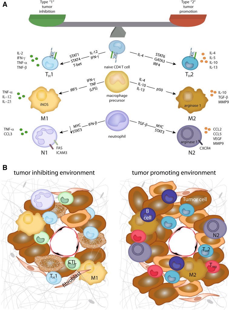
Tumor-inhibiting and tumor-promoting microenvironments. a Depicted are the major factors, and transcription factors involved in the differentiation of type 1 or type 2 cell subtypes. In addition, the main effector molecules from these cells contributing to their tumor-inhibiting or tumor-promoting effect, respectively, are displayed. b In a tumor-inhibiting microenvironment, type 1 cells together with cytotoxic T cells (CTL) contribute to tumor cell destruction. In contrast, in a tumor-promoting microenvironment, type 2 cells are accompanied by regulatory T cells (Treg) and B lymphocytes inhibiting a cytotoxic immune reaction and promoting tumor cell growth, angiogenesis, and metastasis
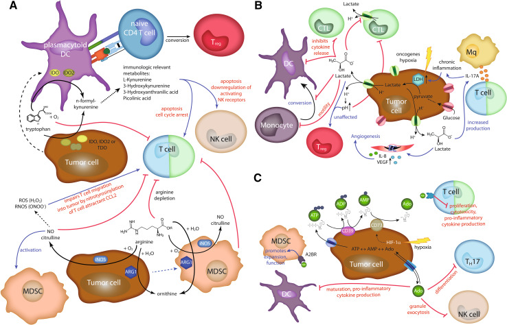
Metabolic enzymes and metabolites in immunosuppression. a Immunosuppression by metabolism of the amino acids tryptophan (upper part) or arginine (lower part). Tumor cells can express the enzymes indoleamine-2,3-dioxygenase (IDO), indoleamine-2,3-dioxygenase-like protein (IDO2), and tryptophan 2,3-dioxygenase (TDO). All these enzymes are able to catalyze the first step in kynurenine pathway of tryptophan metabolism. In addition, plasmacytoid DCs in the draining lymph nodes of cancer patients can express IDO and IDO2. The general depletion of tryptophan impairs T cell proliferation. Furthermore, immune modulatory tryptophan metabolites can induce apoptosis and affect activity of T and NK cells. IDO expressing plasmacytoid DCs can also trigger regulatory T cell (Treg) differentiation. Similarly, arginine depletion by arginase I (ARG1) or inducible nitric oxide synthase (iNOS) impairs T cell activation. iNOS can further suppress immune responses by recruitment and activation of MDSC. NO can be further converted into reactive oxygen species (ROS) and reactive nitrogen and oxygen species (RNOS). The latter can impair T cell migration into the tumor by nitrotyrosinylation of the chemokine CCL2. b Tumor cells primarily rely on glycolysis. One important enzyme involved is LDH converting pyruvate into lactate. Its expression is up-regulated by hypoxia or oncogenes. Lactate is secreted from the cells via monocarboxylate transporters accompanied by H+ transport decreasing the extracellular pH. Lactate affects many different processes like inhibition of cytotoxic T cell responses, causing chronic inflammation via triggering enhanced IL-17A cytokine secretion, or increasing secretion of angiogenic factors by endothelial cells (EC). c Immune suppression by adenosine (Ado). Extracellular ATP is converted by CD39 into ADP and then into AMP. CD73 converts AMP into Ado. Ado can be exported and imported from the tumor cell. HIF-1α enhances CD73 expression. Ado interaction with adenosine A2A receptor (A2AR) on T cells impairs their activity, whereas the binding to adenosine A2B receptor (A2BR) on MDSC promotes their recruitment and function. In addition, Ado inhibits the differentiation of Th17 cells, the activity of NK cells, and the maturation of DCs
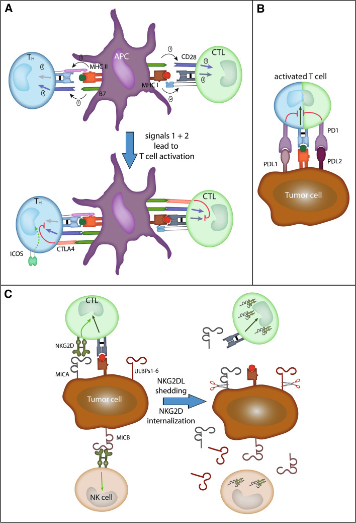
Ligand-mediated lymphocyte control. a Two signals are needed for naive T cell activation: Firstly, CD80 (B7-1) or CD86 (B7-2) has to trigger CD28 signaling, and secondly, the T cell receptor has to interact with the respective peptide/MHC complex. Shortly after activation, CTLA-4 is up-regulated on the T cell-surface competing with CD28 for ligand binding and in turn delivers an inhibitory signal to the T cell. b PD-1 is an inhibitory receptor on activated T cells. Its ligands PDL1 and PDL2 can be expressed on peripheral tissues during inflammation, but also on tumor cells. c In contrast, NKG2D is an activating receptor on NK cells and delivers a co-stimulatory signal to cytotoxic cells (CTLs). Human ligands induced by viral infection, cytokines, or DNA damage are MICA, MICB, and the ULBP1–6. Shedding of these ligands (sNKG2DL) by tumor cells leads to NKG2D receptor internalization thereby preventing their activating effect
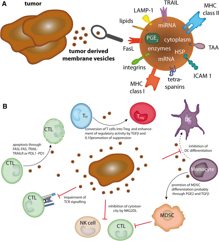
Tumor-derived membrane vesicles. a Molecules present on or in tumor-derived membrane vesicles. b Immune-modulating effects of tumor-derived membrane vesicles (see text for details). CTL cytotoxic lymphocyte, DC dendritic cell, FasL Fas ligand, HSP heat-shock protein, ICAM 1 intercellular adhesion molecule 1, LAMP-1 lysosome-associated membrane protein 1, MDSC myeloid-derived suppressor cell, MHC major histocompatibility complex, miRNA microRNA, mRNA messenger RNA, PD1 programmed cell death protein 1, PDL1 programmed cell death 1 ligand 1, PG prostaglandin, TAA tumor-associated antigen, TCR T cell receptor, T
H T helper cell, T
reg regulatory T cell, TRAIL tumor necrosis factor-related apoptosis-inducing ligand
References
Publication types
MeSH terms
Substances
LinkOut - more resources
Full Text Sources
Other Literature Sources
