Comparative Study
. 2013 Jul;98(7):2887-96.
doi: 10.1210/jc.2012-4000. Epub 2013 May 10.

Expression and function of the progesterone receptor in human prostate stroma provide novel insights to cell proliferation control

Affiliations
Comparative Study

Expression and function of the progesterone receptor in human prostate stroma provide novel insights to cell proliferation control

Yue Yu et al. J Clin Endocrinol Metab. 2013 Jul.

Abstract

Context: Like other tissues, the prostate is an admixture of many different cell types that can be segregated into components of the epithelium or stroma. Reciprocal interactions between these 2 types of cells are critical for maintaining prostate homeostasis, whereas aberrant stromal cell proliferation can disrupt this balance and result in diseases such as benign prostatic hyperplasia. Although the androgen and estrogen receptors are relatively well studied for their functions in controlling stromal cell proliferation and differentiation, the role of the progesterone receptor (PR) remains unclear.

Objective: The aim of the study was to investigate the expression and function of the PR in the prostate.

Design and setting: Human prostate biopsies, renal capsule xenografts, and prostate stromal cells were used. Immunohistochemistry, Western blotting, real-time quantitative PCR, cell proliferation, flow cytometry, and gene microarray analyses were performed.

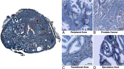
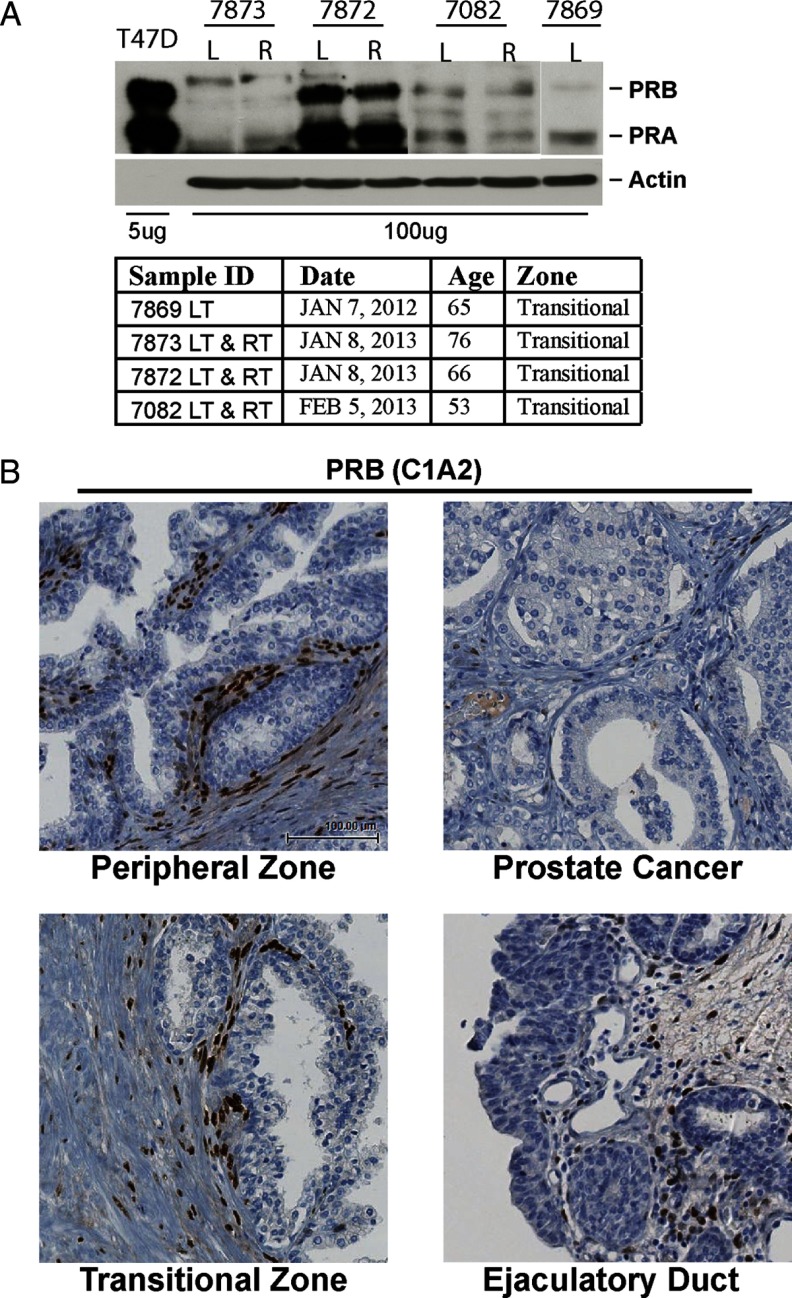
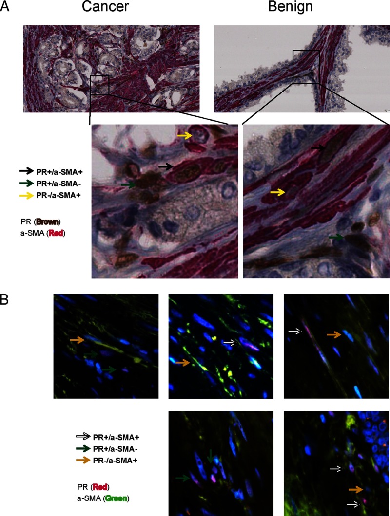
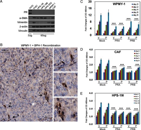
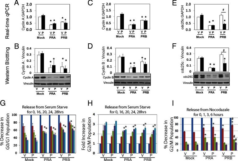
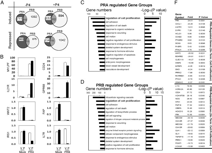
Results: Two PR isoforms, PRA and PRB, are expressed in prostate stromal fibroblasts and smooth muscle cells, but not in epithelial cells. Both PR isoforms suppress prostate stromal cell proliferation through inhibition of the expression of cyclinA, cyclinB, and cdc25c, thus delaying cell cycling through S and M phases. Gene microarray analyses further demonstrated that PRA and PRB regulated different transcriptomes. However, one of the major gene groups commonly regulated by both PR isoforms was the one associated with regulation of cell proliferation.

Conclusion: PR plays an inhibitory role in prostate stromal cell proliferation.


Figures

Figure 1.
Figure 1.
Whole-mount sections of human prostate biopsies (n = 27) were stained with the PR (SP2) antibody by IHC. Four regions within 1 section (ID no. 7219) were amplified: peripheral zone (benign prostate), peripheral zone (cancer prostate), transitional zone, and ejaculatory duct.
Figure 2.
Figure 2.
A, Four prostate tissue samples from transition zones were collected to extract whole cell proteins. Protein extracts (100 μg) were separated on a protein gel together with 5 μg of protein lysates from T47D cells and Western blotted with PR antibody. Information on these 4 tissue samples is also described. B, Whole-mount sections of human prostate biopsies (n = 27) were stained with PRB-specific antibody (C1A2). Four regions within the section (ID no. 7219) were amplified: peripheral zone (benign prostate), peripheral zone (cancer prostate), transitional zone, and ejaculatory duct. LT, left side; RT, right side.
Figure 3.
Figure 3.
A whole-mount section of human prostate tissue (ID no. 7219) was costained with PR and α-SMA antibodies by IHC (A) or by immunofluorescence (B). In IHC studies, PR antibody was detected by UltraMap Anti-Rb HRP, whereas α-SMA antibody was detected by UltraMap Anti-Ms Alk Phos (Ventana Medical Systems, Oro Valley, Arizona). In immunofluorescence studies, PR antibody was detected by Alexa Fluor 568 (Invitrogen, San Diego, California), whereas α-SMA antibody was detected by Alexa Fluor 488 (Invitrogen).
Figure 4.
Figure 4.
A, Whole cell lysates from human prostate stromal cells (60 μg) and T47D breast cancer cells (5 μg) were separated on a protein gel and Western blotted with antibodies against PR, α-SMA, and vimentin. Vinculin and β-actin levels were used as loading controls. B, One million WPMY-1 cells were recombined with 0.5 million BPH-1 cells and grafted under renal capsule in SCID mice for 2 or 4 weeks (n = 3/time point). Xenograft tissues were fixed and immunostained with PR (SP2) antibody. Note that SP2 antibody only recognizes PR protein from human or rat. C and D, WPMY-1 and CAF cells were infected with lentivirus to express mock, PRA, or PRB. Cells were maintained in phenol red free medium containing 10% charcoal stripped serum for 48 hours and then seeded in 96-well culture plates for proliferation assay over 0–8 days. E, Human primary cultured HPS-19I cells were transiently infected with lentivirus encoding mock, PRA, or PRB. Cells were then seeded in 96-well culture plates in the presence of −/+ 10 nm P4. Cell proliferation rates were measured over 0–16 days.
Figure 5.
Figure 5.
A–F, Lentivirus infected WPMY-1 cells expressing mock, PRA, or PRB were maintained in phenol red free medium containing 10% charcoal stripped serum for 48 hours. Cells were treated with either vehicle or 10 nm P4 for 24 hours. Real-time qPCR and Western blotting assays measured mRNA and protein levels of cyclinA, cyclinB, and cdc25c. G and H, Cells were synchronized by serum starvation for 48 hours and then were supplemented with 10% charcoal stripped serum plus −/+10 nm P4 for 0–28 hours. Cells were collected at each time point and subjected to FACS assays. G0/G1 and G2/M cell populations were counted and plotted over the time course. I, Cells were synchronized by 0.1 μg/ml nocodazole for overnight and then released for 0–6 hours. G2/M cell population was counted and plotted over the time course. *One-way ANOVA analyses compared among cells expressing mock, PRA, and PRB with P < .05. #Comparisons between −/+P4 treatments with P < .05.
Figure 6.
Figure 6.
A, Venn diagram showed gene transcription regulated by either PRA or PRB in the presence of −/+ 10 nm P4 from gene microarray analyses. Unpaired t test with P value < 0.05, a fold change > 2.0, and a Benjamini-Hochberg multiple testing correction were applied. B, Validation of 8 representative genes regulated by either PRA or PRB in the presence of −/+ 10 nm P4 was performed by real-time qPCR. C and D, GO analysis on the 5654 genes regulated by either PRA or PRB was performed by using the online tool from DAVID Bioinformatics Resources. Both gene numbers and P values of specific gene groups were listed. F, Within the gene group of regulation of cell proliferation, top-ranked 15 genes regulated by PRA or PRB in the presence of −/+ 10 nm P4 were listed.

References

    1. Chung LW, Davies R. Prostate epithelial differentiation is dictated by its surrounding stroma. Mol Biol Rep. 1996;23:13–19 - PubMed
    1. Cunha GR. Mesenchymal-epithelial interactions: past, present, and future. Differentiation. 2008;76:578–586 - PubMed
    1. Ho CK, Habib FK. Estrogen and androgen signaling in the pathogenesis of BPH. Nat Rev Urol. 2011;8:29–41 - PubMed
    1. Yu S, Yeh CR, Niu Y, et al. Altered prostate epithelial development in mice lacking the androgen receptor in stromal fibroblasts. Prostate. 2012;72:437–449 - PMC - PubMed
    1. Ho CK, Nanda J, Chapman KE, Habib FK. Oestrogen and benign prostatic hyperplasia: effects on stromal cell proliferation and local formation from androgen. J Endocrinol. 2008;197:483–491 - PubMed

Publication types

MeSH terms