Review
. 2013 Jul;53(1):93-102.
doi: 10.1093/icb/ict054. Epub 2013 May 10.

Nuclear magnetic resonance spectroscopy of the circadian clock of cyanobacteria

Affiliations
Review

Nuclear magnetic resonance spectroscopy of the circadian clock of cyanobacteria

Yong-Gang Chang et al. Integr Comp Biol. 2013 Jul.

Abstract

The most well-understood circadian clock at the level of molecular mechanisms is that of cyanobacteria. This overview is on how solution-state nuclear magnetic resonance (NMR) spectroscopy has contributed to this understanding. By exciting atomic spin-½ nuclei in a strong magnetic field, NMR obtains information on their chemical environments, inter-nuclear distances, orientations, and motions. NMR protein samples are typically aqueous, often at near-physiological pH, ionic strength, and temperature. The level of information obtainable by NMR depends on the quality of the NMR sample, by which we mean the solubility and stability of proteins. Here, we use examples from our laboratory to illustrate the advantages and limitations of the technique.


Figures

Fig. 1
Fig. 1
FLAG tags increase stability of the KaiC CI domain. Top panels show 15N, 1H-TROSY spectra of a monomeric variant of the CI domain with a single FLAG tag at the N-terminus before (left panel) and after (right panel) 10-h incubation at 30°C. Bottom panels show spectra of a CI domain having two FLAG tags, one at each terminus. Although a single tag stabilizes CI to a certain extent, two tags had a dramatic effect on long-term7 stability.
Fig. 2
Fig. 2
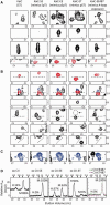
Flexibility of the CII ring depends on the state of phosphorylation at residues S431 and T432 and A-loop position. (A–C) Selected regions from methyl-TROSY spectra of U-[15N, 2H]-Ile-δ1-[13C, 1H]-labeled KaiC (panel 1) and phosphomimics of SpT-KaiC (panel 2), pSpT-KaiC (panel 3), pST-KaiC (panel 4), and pSpT-KaiC487 (panel 5). Horizontal and vertical axes are 1H and 13C chemical shifts in ppm, respectively. Red and blue contours indicate peaks assigned to Ile residues of the CII and CI domains of KaiC, respectively, whereas black contours indicate unassigned peaks. It should be noted that SE-KaiC is ∼30% phosphorylated at residue S431. (D) Gel-filtration profiles of isolated domains of CII (panel 1) and phosphomimics of SpT-CII (panel 2), pSpT-CII (panel 3), pST-CII (panel 4), and pSpT-CII487 and pST-CII487 (panel 5). The hexameric forms of the CI and CII domains eluted at ∼11.7 ml, whereas the monomeric forms eluted at ∼15.4 ml. In panel 5, EE-CII487 and ET-CII487, two KaiC CII domains that were truncated just prior to the A loop at residue 488, eluted as monomers at 16 ml. The percentages of hexameric (H) and monomeric forms (M) are provided in each panel. Adapted from Chang et al. (2011).
Fig. 3
Fig. 3
Profiles of phosphorylation kinetics of KaiC and KaiC phosphomimics. Each data point represents the average of two experiments. The error bars represent the standard error (SEM). (A) Phosphorylation of KaiC in the presence and/or absence of KaiA and KaiB. KaiC alone (circle); KaiC + KaiA (diamond); KaiC + KaiB (triangle); KaiC + KaiA + KaiB (square). (B) Phosphorylation of KaiC variants AT-KaiC (diamond), SE-KaiC (triangle), SA-KaiC (cross), and ET-KaiC (square) in the presence of KaiA. These profiles were obtained by densitometric analysis of SDS–PAGE gels. Adapted from Chang et al. (2011).
Fig. 4
Fig. 4
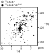
KaiB binds to the CI domain of KaiC. 15N, 1H-HSQC spectra of 15N-labeled KaiB* free (single-contour peaks) and in the presence of CIFLAG (multiple-contour peaks). Adapted from Chang et al. (2012).
Fig. 5
Fig. 5
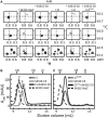
Stacking of CI and CII rings of KaiC is required for binding of KaiC–KaiB. (A) Selected regions from methyl-TROSY spectra of U-[15N, 2H]-Ile-δ1-[13C, 1H]-labeled KaiB alone (panels in column 1) or in the presence of S431E-KaiC (panels in column 2), S431E-CII (panels in column 3), CI (panels in column 4), S431E-CII + CI (panels in column 5), or S431E-CII + CIFLAG (panels in column 6). (B and C) Gel-filtration profiles of mixtures of CI (B) or CIFLAG (C) with S431E-CII. Full-length S431E-KaiC was run as a control for the expected elution position of stacked rings. Compared with CI, CIFLAG has an extra FLAG at its C-terminus, which was engineered for introducing potential repulsive interactions with the N-terminal FLAG tag on isolated S431E-CII domains. Note that the FLAG tag is highly negatively charged “DYKDDDDK”. Adapted from Chang et al. (2012).
Fig. 6
Fig. 6
CII–CI interactions depend on the state of phosphorylation at residues S431 and T432. Selected regions of methyl-TROSY spectra of U-[15N, 2H]-Ile-δ1-[13C, 1H]-labeled KaiC (panel 1) and the phosphomimics of SpT-KaiC (panel 2), pSpT-KaiC (panel 3), pST-KaiC (panel 4), and pSpT-KaiC-487 (panel 5). Horizontal and vertical axes are 1H and 13C chemical shifts in ppm, respectively. All panels were plotted at the same relative contour level. Solid contours are resonances assigned to the CI domain, whereas dashed contours are those unassigned. Selected regions of methyl-TROSY spectra of free CI are shown in panel 6. Adapted from Chang et al. (2011).

Similar articles

References

    1. Akiyama S, Nohara A, Ito K, Maeda Y. Assembly and disassembly dynamics of the cyanobacterial periodosome. Mol Cell. 2008;29:703–16. - PubMed
    1. Amero C, Asuncion Dura M, Noirclerc-Savoye M, Perollier A, Gallet B, Plevin M, Vernet T, Franzetti B, Boisbouvier J. A systematic mutagenesis-driven strategy for site-resolved NMR studies of supramolecular assemblies. J Biomol NMR. 2011;50:229–36. - PubMed
    1. Ayala I, Sounier R, Use N, Gans P, Boisbouvier J. An efficient protocol for the complete incorporation of methyl-protonated alanine in perdeuterated protein. J Biomol NMR. 2009;43:111–9. - PubMed
    1. Barrett PJ, Chen J, Cho M, Kim J, Lu Z, Mathew S, Peng D, Song Y, Van Horn WD, Zhuang T, et al. The quiet renaissance of protein nuclear magnetic resonance. Biochemistry. 2013;52:1303–20. - PMC - PubMed
    1. Bass J. Circadian topology of metabolism. Nature. 2012;491:348–56. - PubMed

Publication types

MeSH terms

Substances

LinkOut - more resources