. 2013 Jun 27;121(26):5238-49.
doi: 10.1182/blood-2012-10-463414. Epub 2013 May 10.

Megakaryocytes promote murine osteoblastic HSC niche expansion and stem cell engraftment after radioablative conditioning

Affiliations

Megakaryocytes promote murine osteoblastic HSC niche expansion and stem cell engraftment after radioablative conditioning

Timothy S Olson et al. Blood. .

Abstract

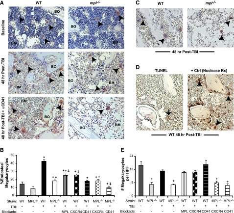
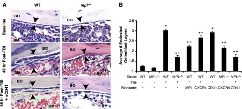
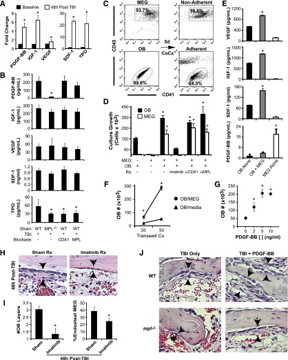
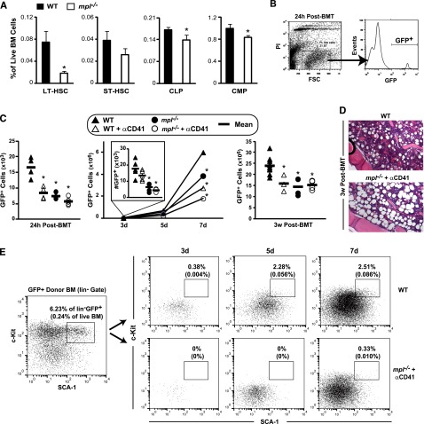
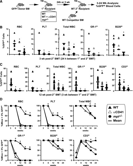
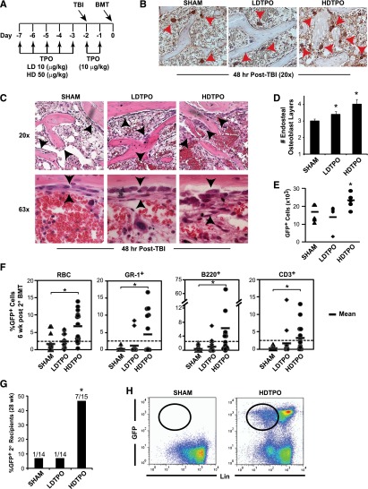
Successful hematopoietic stem cell (HSC) transplantation requires donor HSC engraftment within specialized bone marrow microenvironments known as HSC niches. We have previously reported a profound remodeling of the endosteal osteoblastic HSC niche after total body irradiation (TBI), defined as relocalization of surviving megakaryocytes to the niche site and marked expansion of endosteal osteoblasts. We now demonstrate that host megakaryocytes function critically in expansion of the endosteal niche after preparative radioablation and in the engraftment of donor HSC. We show that TBI-induced migration of megakaryocytes to the endosteal niche depends on thrombopoietin signaling through the c-MPL receptor on megakaryocytes, as well as CD41 integrin-mediated adhesion. Moreover, niche osteoblast proliferation post-TBI required megakaryocyte-secreted platelet-derived growth factor-BB. Furthermore, blockade of c-MPL-dependent megakaryocyte migration and function after TBI resulted in a significant decrease in donor HSC engraftment in primary and competitive secondary transplantation assays. Finally, we administered thrombopoietin to mice beginning 5 days before marrow radioablation and ending 24 hours before transplant to enhance megakaryocyte function post-TBI, and found that this strategy significantly enhanced donor HSC engraftment, providing a rationale for improving hematopoietic recovery and perhaps overall outcome after clinical HSC transplantation.


Figures

Figure 1
Figure 1
Megakaryocyte migration to the endosteal niche is induced by TBI and inhibited by c-MPL deficiency and CD41 blockade. (A) Immunostaining of metaphyseal bone (BO) and BM sections (20×) for CD41-expressing (red) megakaryocytes (black arrowheads) from WT (left column) and mpl−/− mice (right column) at baseline (top) and at 48 hours post-TBI in the presence (bottom) or absence (middle) of CD41 blockade. (B) Percentage of endosteal megakaryocytes (mean ± SEM) at baseline (no TBI) or 48 hours after TBI in WT and mpl−/− mice receiving TBI only, anti-c-MPL–blocking antibody (MPL), CXCR4 blockade with AMD3100 (CXCR4), or anti-CD41–blocking antibody (CD41) (n ≥4 mice per group). *P < .05 vs unirradiated group within same strain, +P < .01 vs WT mice receiving TBI only in other TBI-treated groups, ‡P < .05 vs mpl−/− mice receiving TBI only in groups with specific receptor blockade (comparisons performed by 1-way ANOVA and Dunnett posttest analysis). (C) Immunostaining (20×) for BrdU incorporation (black) in CD41-expressing (red) megakaryocytes (arrowheads) in WT vs mpl−/− BM 48 hours after TBI. (D) At 48 hours post-TBI in WT BM (left), few BM cells remain, and none with the morphologic appearance of megakaryocytes display TUNEL+ (brown stain, black arrow) apoptosis, compared with a ligase-treated positive control serial section (right), with megakaryocytes (arrowheads) present in each section (20×). (E) Total megakaryocyte number per HPF (20×) in the same groups as in (B). +P < .01 vs baseline WT or WT TBI-only group (comparisons performed by 1-way ANOVA and Dunnett posttest analysis).
Figure 2
Figure 2
Blockade of TBI-induced c-MPL–dependent megakaryocyte endosteal migration abrogates niche osteoblast expansion post-TBI. (A) H&E sections (63×) of WT (left panels) and mpl−/− (right panels) metaphyseal bone (BO) and BM at baseline (top) and 48 hours post-TBI in the presence (bottom) or absence (middle) of CD41 blockade, demonstrating a single layer of endosteal osteoblasts at baseline followed by proliferation and expansion of endosteal osteoblasts in WT BM 48 hours post-TBI that is markedly attenuated by c-MPL deficiency, but not CD41 blockade. (B) Quantitative scoring analysis of osteoblast layer number (mean ± SEM) at baseline in WT and mpl−/− mice (n = 5 mice each); in WT mice receiving TBI only (n = 19), or blockade of c-MPL (n = 10), CXCR4, or CD41 (n = 5 each); and in mpl−/− mice receiving TBI only (n = 19) or TBI plus blockade of CXCR4 or CD41 (n = 5 each). *P < .01 vs unirradiated group within same strain, +P < .05 vs WT mice receiving TBI only in TBI-treated groups (comparisons performed by 1-way ANOVA and Dunnett posttest analysis).
Figure 3
Figure 3
Megakaryocytes drive osteoblast expansion through PDGF-BB expression. (A) BM levels of PDGF-BB, IGF-1, VEGF, SDF-1, and TPO by ELISA (mean ± SD) at baseline and at 48 hours post-TBI in WT mice. *P < .001 vs baseline. (B) Levels of BM PDGF-BB, IGF-1, VEGF, SDF-1, and levels in WT vs mpl−/− mice with or without CD41 or c-MPL antibody blockade (n = 5 per group) at 48 hours post-TBI. *P < .05 vs WT TBI-only group. (C) Representative dot plots showing purity of isolated, ex vivo–expanded megakaryocytes (MEG) and primary osteoblasts (OB) before coculture (left), together with nonadherent (top right) MEG and adherent (bottom right) OB fractions after 5 days of coculture. (D) Five-day growth/survival of MEG and OB cultured separately or in combination (OB+MEG), with or without imatinib (5 μm), anti-CD41 antibody, or anti-c-MPL antibody (n = 3 each; mean ± SD). *P < .01 vs OB cultured alone, +P < .001 vs MEG cultured alone. (E) VEGF, IGF-1, SDF-1, and PDGF-BB supernatant concentrations (mean ± SD, n = 3 each) from 5-day cultures of OB, MEG, or cocultured OB + MEG. *P < .001 vs OB or MEG alone, +P < .001 vs OB alone or OB + MEG. (F) OB growth in 2- and 5-day OB cultures in which either MEG or medium alone was added to cultures across a 0.4-µM transwell membrane (n = 3 each). *P < .002 vs OB cultured with medium alone. (G) Five-day cultures of OB cultured with media alone or with increasing concentrations (2-10 ng/mL) of recombinant PDGF-BB (n = 3 each). *P < .01 vs cultures without PDGF-BB. (H) H&E-stained sections (40×) of BM at 48 hours post-TBI, demonstrating abrogation of OB expansion (arrowheads) caused by imatinib (right) vs sham (left) treatment. (I) Quantitative analysis of average OB layers (left) and percentage of endosteal MEG (right) at 48 hours post-TBI in imatinib vs sham-treated WT mice (n = 5 mice per group). *P < .005 vs sham-treated mice. (J) 40× photomicrographs of H&E-stained metaphyseal BM sections at 48 hours post-TBI in WT and mpl−/− mice demonstrating that treatment with PDGF-BB enhances 48-hour post-TBI endosteal OB proliferation (black arrows) in both WT and mpl−/− BM.
Figure 4
Figure 4
Blockade of host megakaryocyte function post-TBI decreases BM engraftment and expansion in primary transplant recipients. (A) Baseline LT-HSC (c-kit+LinSCA-1+CD135lo), ST-HSC (c-kit+LinSCA-1+CD135+CD34+), common lymphoid progenitors (CLP, LinCD127+), and common myeloid progenitors (CMP, c-kit+LinSCA-1CD34+CD127) in WT and mpl−/− BM, expressed as the percentage of all live BM cells (mean ± SD, n = 3 each). *P < .05 vs percentage in WT mice. (B) Representative dot plot and histogram demonstrating method of determining percentages of live, GFP+ donor cells within irradiated recipient BM at 24 hours after BMT, used with total BM cell counts to determine absolute donor GFP+ BM cells. (C) Absolute live BM GFP+ donor cells (individual mice and/or mean per group) shown at 24 hours (left), 3 to 7 days (middle), and 3 weeks (right) post-BMT of 5 × 10⁶ (24 hours to 7 days), or 2 × 10⁵ (3 weeks) GFP+ whole BM cells into post-TBI primary recipient WT or mpl−/− mice ± recipient treatment with anti-CD41 at the time of TBI (n = 3-5 mice per group). *P < .05 vs WT recipients. (D) H&E-stained histologic sections showing decreased BM cellularity in anti-CD41–treated mpl−/− (bottom) vs WT (top) recipients of GFP+ donor BM at 3 weeks post-BMT. (E) Flow cytometry dot plots of c-Kit vs SCA-1 expression on live GFP+Lin gated whole BM cells from H2K-GFP donor BM before transplant (used to establish the HSC/progenitor c-Kit+linSca-1+ [KLS] gate), and from either WT (top, right) or anti-CD41–treated mpl−/− (bottom, right) primary recipients (pooled BM from 2-4 mice per time point), at 3, 5, and 7 days post-BMT with GFP+ whole BM cells (3-5 × 10⁶ cells per mouse). Rectangle in each plot represents KLS population with frequency expressed as a percentage of live GFP+Lin cells and of total live cells (in parentheses).
Figure 5
Figure 5
LT-HSC engraftment after BMT is severely impaired by blockade of host megakaryocytes. (A) GFP+ donor engraftment in competitive secondary (2°) transplant (BMT) assays in which GFP+ donor BM was transplanted (5 × 10⁶ BM cells) into post-TBI WT, anti-CD41–treated WT, and mpl−/− primary (1°) recipients. (B) In the initial assay, BM was harvested 24 hours after 1° BMT and transplanted with 2 × 10⁵ WT competitor BM cells into post-TBI WT 2° recipients. GFP+ peripheral RBC, platelet, total WBC, myeloid (GR-1+), and B-cell (B220+) engraftment at 3 weeks after 2° BMT is shown. *P < .05 vs 2° recipients receiving WT 1° recipient BM. (C) Separate competitive transplant assay showing GFP+ peripheral RBC, platelet, total WBC, myeloid, B-cell, and T-cell (CD3+) engraftment at 12 weeks after competitive 2° BMT with 105 WT competitor BM cells, and either WT, WT + αCD41, or mpl−/− 1° recipient BM, harvested 3 weeks after 1° recipient BMT with 2 × 10⁵ GFP+ donor BM cells. *P < .05 vs 2° recipients receiving WT 1° recipient cells. (D) Percentage of competitively transplanted 2° recipients receiving WT, WT + αCD41, or mpl−/− 1° recipients cells 3 weeks after 1° BMT demonstrating >3% GFP+ cells in specified lineage from 3 to 24 weeks after 2° BMT. *P < .05 vs percentage of 2° recipients of anti-CD41–treated or mpl−/− 1° recipient BM.
Figure 6
Figure 6
TPO administration before BMT enhances megakaryocyte-mediated expansion of niche osteoblasts and HSC engraftment. (A) Schedule of TPO administration. (B) Immunostained (CD41) sections (20×) comparing 48-hour post-TBI BM endosteal megakaryocytes (red arrowheads) after high-dose TPO (HDTPO, 50 µg/kg), low-dose TPO (LDTPO, 10 µg/kg), or sham treatment. (C) BM sections (20× top, 63× bottom) at 48 hours post-TBI showing increased endosteal osteoblast expansion (black arrowheads) after HDTPO treatment vs LDTPO- or sham-treated mice. (D) Quantitative assessment of 48-hour post-TBI osteoblast layers (mean ± SD) in sham-, LDTPO-, and HDTPO-treated WT mice (n ≥5 mice per group). *P < .005 vs sham-treated group. (E) Absolute BM GFP+ engraftment at 24 hours post-BMT (5 × 10⁶ cells) in post-TBI 1° recipients receiving sham, LDTPO, or HDTPO treatment (n = 5 per group). *P < .05 vs sham-treated group. (F) Secondary (2°) recipient GFP+ peripheral RBC, myeloid (GR-1+), B-cell (B220+), and T-cell (CD3+) engraftment at 6 weeks after 2° BMT with 2 × 10⁵ WT competitor BM cells and either sham-, LDTPO-, or HDTPO-treated primary (1°) recipient BM at 24 hours after 1° recipient BMT. *P < .02 vs 2° recipients of sham-treated 1° recipient BM. (G) Percentage of 2° recipients of sham, LDTPO, or HDTPO 1° recipient BM with GFP+ BM engraftment (>0.25% each) in myeloid, B-cell, T-cell, and Lin- BM populations at 28 weeks after 2° BMT. *P < .05 vs 2° recipients of sham-treated 1° recipient BM. (H) Representative dot plots of GFP vs Lin in BM at 28 weeks after 2° BMT in recipients of sham- vs HDTPO-treated 1° recipient BM.

Similar articles

Cited by

References

    1. Escalón MP, Komanduri KV. Cord blood transplantation: evolving strategies to improve engraftment and immune reconstitution. Curr Opin Oncol. 2010;22(2):122–129. - PubMed
    1. de la Morena MT, Gatti RA. A history of bone marrow transplantation. Immunol Allergy Clin North Am. 2010;30(1):1–15. - PubMed
    1. Garrett RW, Emerson SG. Bone and blood vessels: the hard and the soft of hematopoietic stem cell niches. Cell Stem Cell. 2009;4(6):503–506. - PubMed
    1. Zhang J, Niu C, Ye L, et al. Identification of the haematopoietic stem cell niche and control of the niche size. Nature. 2003;425(6960):836–841. - PubMed
    1. Xie Y, Yin T, Wiegraebe W, et al. Detection of functional haematopoietic stem cell niche using real-time imaging. Nature. 2009;457(7225):97–101. - PubMed

Publication types

MeSH terms