

Fatigue and non-fatigue mathematical muscle models during functional electrical stimulation of paralyzed muscle
- PMID: 23667385
- PMCID: PMC3647619
- DOI: 10.1016/j.bspc.2009.12.001
Fatigue and non-fatigue mathematical muscle models during functional electrical stimulation of paralyzed muscle
Abstract
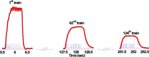
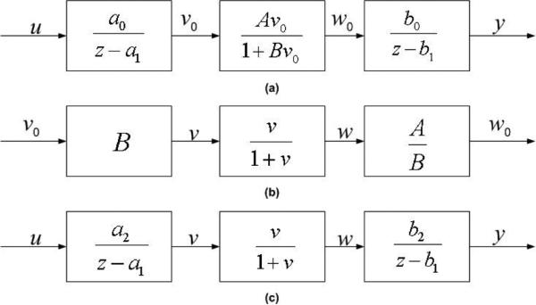
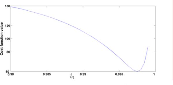
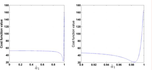
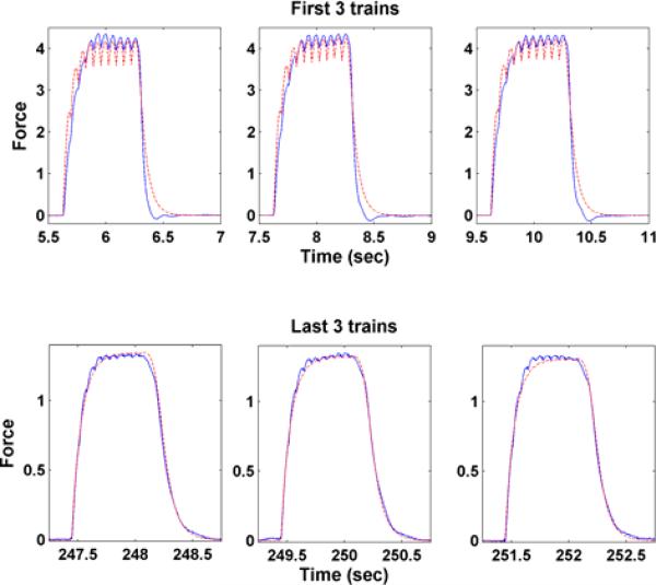
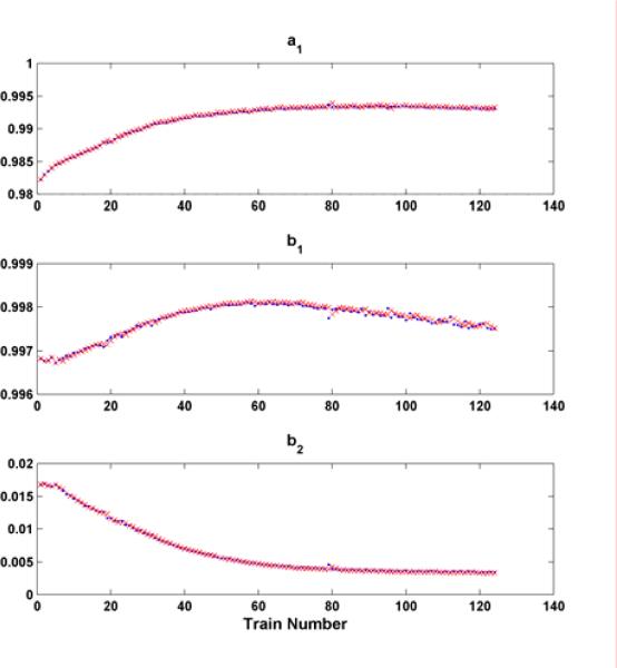
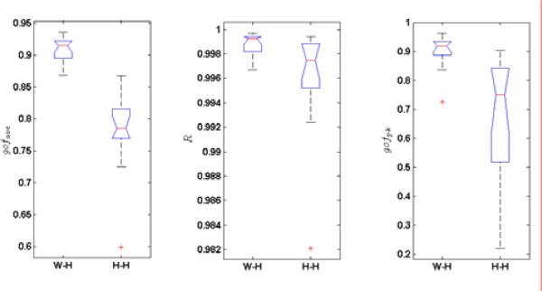
Electrical muscle stimulation demonstrates potential for preventing muscle atrophy and for restoring functional movement after spinal cord injury (SCI). Control systems used to optimize delivery of electrical stimulation protocols depend upon the algorithms generated using computational models of paralyzed muscle force output. The Hill-Huxley-type model, while being highly accurate, is also very complex, making it difficult for real-time implementation. In this paper, we propose a Wiener-Hammerstein system to model the paralyzed skeletal muscle under electrical stimulus conditions. The proposed model has substantial advantages in identification algorithm analysis and implementation including computational complexity and convergence, which enable it to be used in real-time model implementation. Experimental data sets from the soleus muscles of fourteen subjects with SCI were collected and tested. The simulation results show that the proposed model outperforms the Hill-Huxley-type model not only in peak force prediction, but also in fitting performance for force output of each individual stimulation train.
Figures

Similar articles
-
Identification of a Modified Wiener-Hammerstein System and Its Application in Electrically Stimulated Paralyzed Skeletal Muscle Modeling.Automatica (Oxf). 2009 Mar;45(3):736-743. doi: 10.1016/j.automatica.2008.09.023. Automatica (Oxf). 2009. PMID: 23467426 Free PMC article.
-
Mathematical models of human paralyzed muscle after long-term training.J Biomech. 2007;40(12):2587-95. doi: 10.1016/j.jbiomech.2006.12.015. Epub 2007 Feb 20. J Biomech. 2007. PMID: 17316653 Free PMC article. Clinical Trial.
-
Doublet electrical stimulation enhances torque production in people with spinal cord injury.Neurorehabil Neural Repair. 2011 Jun;25(5):423-32. doi: 10.1177/1545968310390224. Epub 2011 Feb 8. Neurorehabil Neural Repair. 2011. PMID: 21304018 Free PMC article.
-
A review of methods for achieving upper limb movement following spinal cord injury through hybrid muscle stimulation and robotic assistance.Exp Neurol. 2020 Jun;328:113274. doi: 10.1016/j.expneurol.2020.113274. Epub 2020 Mar 5. Exp Neurol. 2020. PMID: 32145251 Review.
-
Electrical stimulation for therapy and mobility after spinal cord injury.Prog Brain Res. 2002;137:27-34. doi: 10.1016/s0079-6123(02)37005-5. Prog Brain Res. 2002. PMID: 12440357 Review.
Cited by
-
Evoked Electromyographically Controlled Electrical Stimulation.Front Neurosci. 2016 Jul 14;10:335. doi: 10.3389/fnins.2016.00335. eCollection 2016. Front Neurosci. 2016. PMID: 27471448 Free PMC article.
-
Exhaustion of Skeletal Muscle Fibers Within Seconds: Incorporating Phosphate Kinetics Into a Hill-Type Model.Front Physiol. 2020 May 5;11:306. doi: 10.3389/fphys.2020.00306. eCollection 2020. Front Physiol. 2020. PMID: 32431619 Free PMC article.
-
Design of the Cooperative Actuation in Hybrid Orthoses: A Theoretical Approach Based on Muscle Models.Front Neurorobot. 2019 Jul 31;13:58. doi: 10.3389/fnbot.2019.00058. eCollection 2019. Front Neurorobot. 2019. PMID: 31417390 Free PMC article.
-
A Pilot Study on EEG-Based Evaluation of Visually Induced Motion Sickness.J Imaging Sci Technol. 2020 Mar 1;64(2):205011-2050110. doi: 10.2352/J.ImagingSci.Technol.2020.64.2.020501. Epub 2020 Jan 31. J Imaging Sci Technol. 2020. PMID: 33907364 Free PMC article.
References
-
- Castro MJ, Apple DF, Hillegass EA, Dudley GA. Influence of complete spinal cord injury on skeletal muscle cross-sectional area within the first 6 months of injury. European Journal of Applied Physiology & Occupational Physiology. 1999;86(1):350–358. - PubMed
-
- Vestergaard P, Krogh K, Rejnmark L, Mosekilde L. Fracture rates and risk factors for fractures in patients with spinal cord injury. Spinal Cord. 1998;36(11):790–796. - PubMed
-
- Mahoney ET, Bickel CS, Elder C, Black C, Slade JM, Apple D, Dudley GA. Changes in skeletal muscle size and glucose tolerance with electrically stimulated resistance training in subjects with chronic spinal cord injury. Arch. Phys. Med. Rehabil. 2005;86(7):1502–1504. - PubMed
-
- Dudley GA, Castro MJ, Rogers S, Apple DF. A simple means of increasing muscle size after spinal cord injury: a pilot study. European Journal of Applied Physiology & Occupational Physiology. 1999;80(4):394–396. - PubMed
Grants and funding
LinkOut - more resources
Full Text Sources