

Tetramerization reinforces the dimer interface of MnSOD
- PMID: 23667478
- PMCID: PMC3646814
- DOI: 10.1371/journal.pone.0062446
Tetramerization reinforces the dimer interface of MnSOD
Abstract
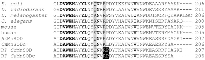
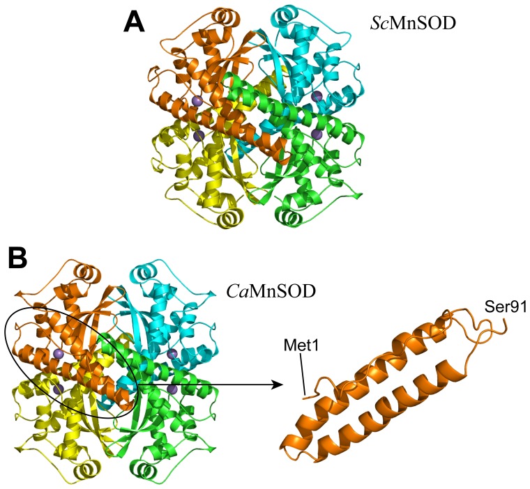
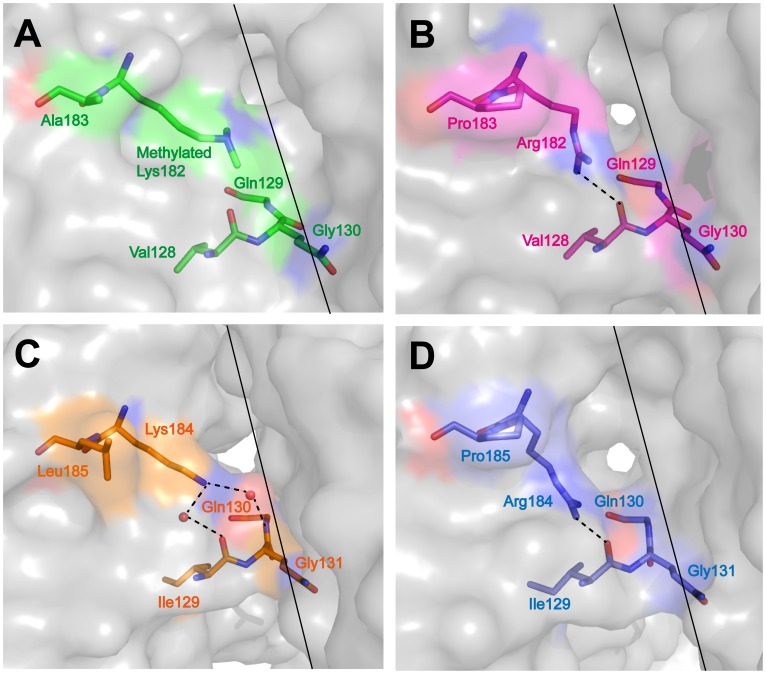
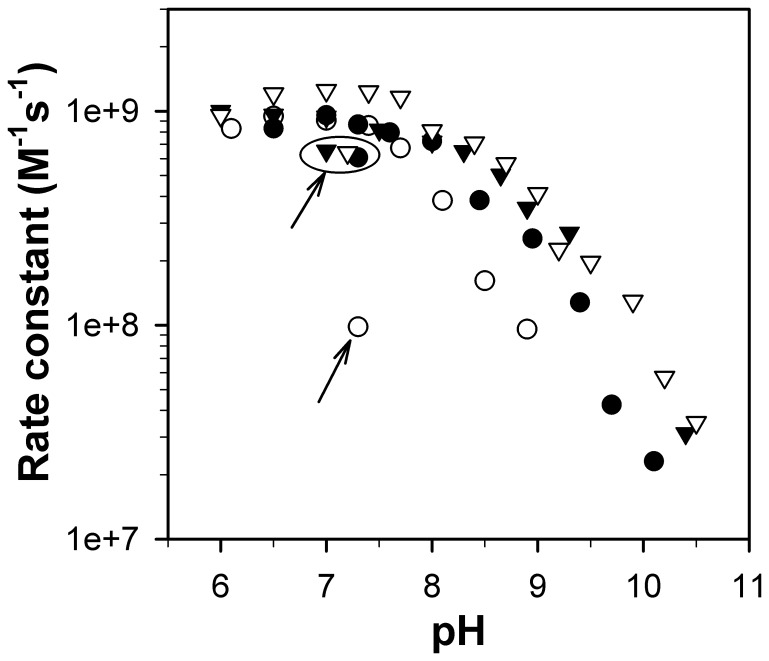
Two yeast manganese superoxide dismutases (MnSOD), one from Saccharomyces cerevisiae mitochondria (ScMnSOD) and the other from Candida albicans cytosol (CaMnSODc), have most biochemical and biophysical properties in common, yet ScMnSOD is a tetramer and CaMnSODc is a dimer or "loose tetramer" in solution. Although CaMnSODc was found to crystallize as a tetramer, there is no indication from the solution properties that the functionality of CaMnSODc in vivo depends upon the formation of the tetrameric structure. To elucidate further the functional significance of MnSOD quaternary structure, wild-type and mutant forms of ScMnSOD (K182R, A183P mutant) and CaMnSODc (K184R, L185P mutant) with the substitutions at dimer interfaces were analyzed with respect to their oligomeric states and resistance to pH, heat, and denaturant. Dimeric CaMnSODc was found to be significantly more subject to thermal or denaturant-induced unfolding than tetrameric ScMnSOD. The residue substitutions at dimer interfaces caused dimeric CaMnSODc but not tetrameric ScMnSOD to dissociate into monomers. We conclude that the tetrameric assembly strongly reinforces the dimer interface, which is critical for MnSOD activity.
Conflict of interest statement
Figures
Similar articles
-
Comparison of two yeast MnSODs: mitochondrial Saccharomyces cerevisiae versus cytosolic Candida albicans.J Am Chem Soc. 2011 Dec 28;133(51):20878-89. doi: 10.1021/ja2077476. Epub 2011 Nov 30. J Am Chem Soc. 2011. PMID: 22077216 Free PMC article.
-
Biophysical characterization of the dimer and tetramer interface interactions of the human cytosolic malic enzyme.PLoS One. 2012;7(12):e50143. doi: 10.1371/journal.pone.0050143. Epub 2012 Dec 21. PLoS One. 2012. PMID: 23284632 Free PMC article.
-
Human mitochondrial manganese superoxide dismutase polymorphic variant Ile58Thr reduces activity by destabilizing the tetrameric interface.Biochemistry. 1996 Apr 9;35(14):4287-97. doi: 10.1021/bi951892w. Biochemistry. 1996. PMID: 8605177
-
Role of a glutamate bridge spanning the dimeric interface of human manganese superoxide dismutase.Biochemistry. 2008 Apr 22;47(16):4621-8. doi: 10.1021/bi7024518. Epub 2008 Mar 29. Biochemistry. 2008. PMID: 18373354
-
Investigation of the highly active manganese superoxide dismutase from Saccharomyces cerevisiae.J Am Chem Soc. 2010 Sep 15;132(36):12525-7. doi: 10.1021/ja104179r. J Am Chem Soc. 2010. PMID: 20726524 Free PMC article.
Cited by
-
Redox manipulation of the manganese metal in human manganese superoxide dismutase for neutron diffraction.Acta Crystallogr F Struct Biol Commun. 2018 Oct 1;74(Pt 10):677-687. doi: 10.1107/S2053230X18011299. Epub 2018 Sep 21. Acta Crystallogr F Struct Biol Commun. 2018. PMID: 30279321 Free PMC article.
-
Superoxide dismutases and superoxide reductases.Chem Rev. 2014 Apr 9;114(7):3854-918. doi: 10.1021/cr4005296. Epub 2014 Apr 1. Chem Rev. 2014. PMID: 24684599 Free PMC article. Review. No abstract available.
-
Unique Characteristics of Recombinant Hybrid Manganese Superoxide Dismutase from Staphylococcus equorum and S. saprophyticus.Protein J. 2016 Apr;35(2):136-44. doi: 10.1007/s10930-016-9650-5. Protein J. 2016. PMID: 26960678
-
Manganese Superoxide Dismutase Acetylation and Regulation of Protein Structure in Breast Cancer Biology and Therapy.Antioxidants (Basel). 2022 Mar 25;11(4):635. doi: 10.3390/antiox11040635. Antioxidants (Basel). 2022. PMID: 35453320 Free PMC article. Review.
-
A Study of the Activity of Recombinant Mn-Superoxide Dismutase in the Presence of Gold and Silver Nanoparticles.Appl Biochem Biotechnol. 2019 Apr;187(4):1551-1568. doi: 10.1007/s12010-018-2896-y. Epub 2018 Oct 3. Appl Biochem Biotechnol. 2019. PMID: 30284207 Free PMC article.
References
-
- Wagner UG, Pattridge KA, Ludwig ML, Stallings WC, Werber MM, et al. (1993) Comparison of the crystal structures of genetically engineered human manganese superoxide dismutase and manganese superoxide dismutase from Thermus thermophilus: differences in dimer-dimer interaction. Protein Sci 2: 814–825. - PMC - PubMed
-
- Edwards RA, Baker HM, Whittaker MM, Whittaker JW, Jameson GB, et al. (1998) Crystal structure of Escherichia coli manganese superoxide dismutase at 2.1-angstrom resolution. Journal of Biological Inorganic Chemistry 3: 161–171.
-
- Abreu IA, Hearn A, An H, Nick HS, Silverman DN, et al. (2008) The kinetic mechanism of manganese-containing superoxide dismutase from Deinococcus radiodurans: a specialized enzyme for the elimination of high superoxide concentrations. Biochemistry 47: 2350–2356. - PubMed
-
- Borgstahl GE, Parge HE, Hickey MJ, Beyer WF Jr, Hallewell RA, et al. (1992) The structure of human mitochondrial manganese superoxide dismutase reveals a novel tetrameric interface of two 4-helix bundles. Cell 71: 107–118. - PubMed
Publication types
MeSH terms
Substances
Grants and funding
LinkOut - more resources
Full Text Sources
Other Literature Sources
Molecular Biology Databases
