

Stanniocalcin-1 protects retinal ganglion cells by inhibiting apoptosis and oxidative damage
- PMID: 23667669
- PMCID: PMC3646795
- DOI: 10.1371/journal.pone.0063749
Stanniocalcin-1 protects retinal ganglion cells by inhibiting apoptosis and oxidative damage
Abstract
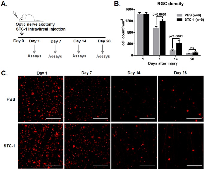
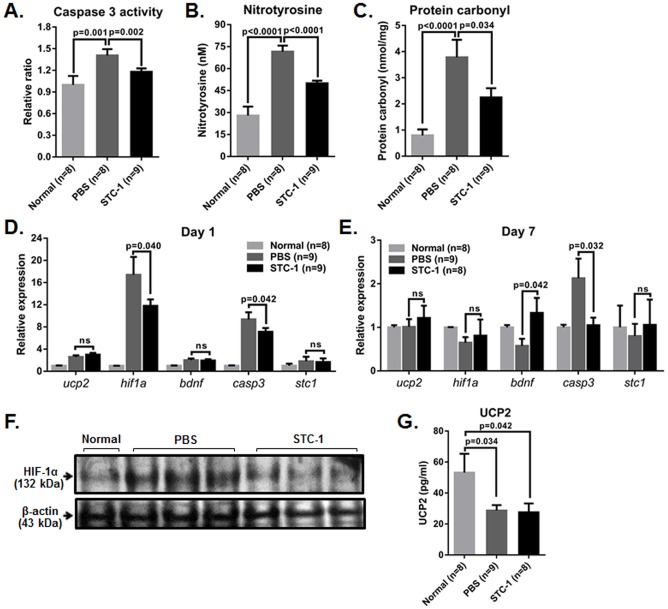
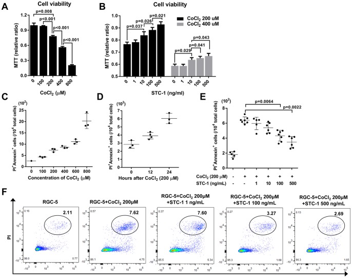
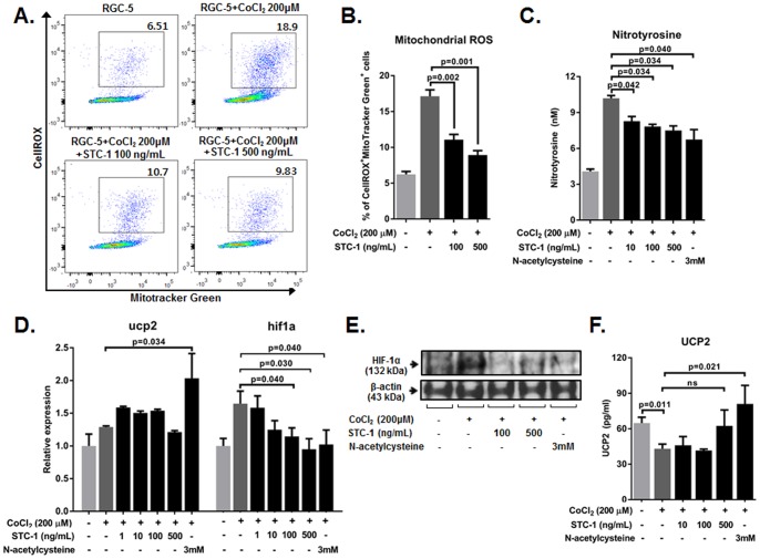
Optic neuropathy including glaucoma is one of the leading causes of irreversible vision loss, and there are currently no effective therapies. The hallmark of pathophysiology of optic neuropathy is oxidative stress and apoptotic death of retinal ganglion cells (RGCs), a population of neurons in the central nervous system with their soma in the inner retina and axons in the optic nerve. We here tested that an anti-apoptotic protein stanniocalcin-1 (STC-1) can prevent loss of RGCs in the rat retina with optic nerve transection (ONT) and in cultures of RGC-5 cells with CoCl2 injury. We found that intravitreal injection of STC-1 increased the number of RGCs in the retina at days 7 and 14 after ONT, and decreased apoptosis and oxidative damage. In cultures, treatment with STC-1 dose-dependently increased cell viability, and decreased apoptosis and levels of reactive oxygen species in RGC-5 cells that were exposed to CoCl2. The expression of HIF-1α that was up-regulated by injury was significantly suppressed in the retina and in RGC-5 cells by STC-1 treatment. The results suggested that intravitreal injection of STC-1 might be a useful therapy for optic nerve diseases in which RGCs undergo apoptosis through oxidative stress.
Conflict of interest statement
Figures
Similar articles
-
Intravitreal delivery of human NgR-Fc decoy protein regenerates axons after optic nerve crush and protects ganglion cells in glaucoma models.Invest Ophthalmol Vis Sci. 2015 Feb 5;56(2):1357-66. doi: 10.1167/iovs.14-15472. Invest Ophthalmol Vis Sci. 2015. PMID: 25655801 Free PMC article.
-
Dimercaprol attenuates oxidative stress-induced damage of retinal ganglion cells in an in vitro and in vivo model of traumatic optic neuropathy.Neuropharmacology. 2025 Oct 1;277:110525. doi: 10.1016/j.neuropharm.2025.110525. Epub 2025 May 21. Neuropharmacology. 2025. PMID: 40409536
-
Rotenone-Induced Optic Nerve Damage and Retinal Ganglion Cell Loss in Rats.Biomolecules. 2024 Aug 23;14(9):1047. doi: 10.3390/biom14091047. Biomolecules. 2024. PMID: 39334813 Free PMC article.
-
The molecular basis of retinal ganglion cell death in glaucoma.Prog Retin Eye Res. 2012 Mar;31(2):152-81. doi: 10.1016/j.preteyeres.2011.11.002. Epub 2011 Dec 4. Prog Retin Eye Res. 2012. PMID: 22155051 Review.
-
Neuroprotective effects of resveratrol on retinal ganglion cells in glaucoma in rodents: A narrative review.Animal Model Exp Med. 2024 Jun;7(3):195-207. doi: 10.1002/ame2.12438. Epub 2024 May 29. Animal Model Exp Med. 2024. PMID: 38808561 Free PMC article. Review.
Cited by
-
Investigating a downstream gene of Gpnmb using the systems genetics method.Mol Vis. 2019 Apr 23;25:222-236. eCollection 2019. Mol Vis. 2019. PMID: 31057322 Free PMC article.
-
AAV-mediated STC-1 expression mitigates neuroinflammation and preserves visual function in degenerative retinopathy.J Transl Med. 2025 Aug 18;23(1):924. doi: 10.1186/s12967-025-06898-1. J Transl Med. 2025. PMID: 40826460 Free PMC article.
-
Stanniocalcin 2 enhances mesenchymal stem cell survival by suppressing oxidative stress.BMB Rep. 2015 Dec;48(12):702-7. doi: 10.5483/bmbrep.2015.48.12.158. BMB Rep. 2015. PMID: 26424558 Free PMC article.
-
CD200Fc Attenuates Retinal Glial Responses and RGCs Apoptosis After Optic Nerve Crush by Modulating CD200/CD200R1 Interaction.J Mol Neurosci. 2018 Feb;64(2):200-210. doi: 10.1007/s12031-017-1020-z. Epub 2017 Dec 26. J Mol Neurosci. 2018. PMID: 29280053
-
Stanniocalcin-1 Protects a Mouse Model from Renal Ischemia-Reperfusion Injury by Affecting ROS-Mediated Multiple Signaling Pathways.Int J Mol Sci. 2016 Jul 12;17(7):1051. doi: 10.3390/ijms17071051. Int J Mol Sci. 2016. PMID: 27420048 Free PMC article.
References
-
- Fischer D, Leibinger M (2012) Promoting optic nerve regeneration. Prog Retin Eye Res 31: 688–701. - PubMed
-
- Almasieh M, Wilson AM, Morquette B, Cueva Vargas JL, Di Polo A (2012) The molecular basis of retinal ganglion cell death in glaucoma. Prog Retin Eye Res 31: 152–181. - PubMed
-
- Andersen JK (2004) Oxidative stress in neurodegeneration: cause or consequence? Nat Med 5: S18–S25. - PubMed
-
- Nguyen SM, Alexejun CN, Levin LA (2003) Amplification of a reactive oxygen species signal in axotomized retinal ganglion cells. Antioxid Redox Signal 5: 629–634. - PubMed
Publication types
MeSH terms
Substances
LinkOut - more resources
Full Text Sources
Other Literature Sources
