. 2013 Jun 25;7(6):4855-68.
doi: 10.1021/nn305872d. Epub 2013 May 23.

Cellular interaction and toxicity depend on physicochemical properties and surface modification of redox-active nanomaterials

Affiliations

Cellular interaction and toxicity depend on physicochemical properties and surface modification of redox-active nanomaterials

Janet M Dowding et al. ACS Nano. .

Abstract

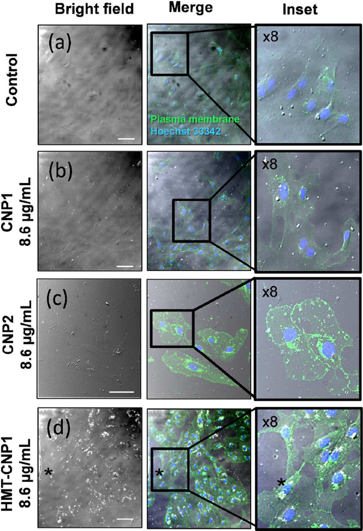
The study of the chemical and biological properties of CeO2 nanoparticles (CNPs) has expanded recently due to its therapeutic potential, and the methods used to synthesize these materials are diverse. Moreover, conflicting reports exist regarding the toxicity of CNPs. To help resolve these discrepancies, we must first determine whether CNPs made by different methods are similar or different in their physicochemical and catalytic properties. In this paper, we have synthesized several forms of CNPs using identical precursors through a wet chemical process but using different oxidizer/reducer; H2O2 (CNP1), NH4OH (CNP2), or hexamethylenetetramine (HMT-CNP1). Physicochemical properties of these CNPs were extensively studied and found to be different depending on the preparation methods. Unlike CNP1 and CNP2, HMT-CNP1 was readily taken into endothelial cells and the aggregation can be visualized using light microscopy. Exposure to HMT-CNP1 also reduced cell viability at a 10-fold lower concentration than CNP1 or CNP2. Surprisingly, exposure to HMT-CNP1 led to substantial decreases in ATP levels. Mechanistic studies revealed that HMT-CNP1 exhibited substantial ATPase (phosphatase) activity. Though CNP2 also exhibits ATPase activity, CNP1 lacked ATPase activity. The difference in catalytic (ATPase) activity of different CNPs preparation may be due to differences in their morphology and oxygen extraction energy. These results suggest that the combination of increased uptake and ATPase activity of HMT-CNP1 may underlie the biomechanism of the toxicity of this preparation of CNPs and may suggest that ATPase activity should be considered when synthesizing CNPs for use in biomedical applications.


Figures

Figure 1
Figure 1
Size, shape, and morphology variation of Cerium Oxide Nanoparticles (CeO2) NPs synthesized by two different synthesis methods. TEM images of CeO2 NPs prepared using water-based (a & b) or solvent HMT (c – e) synthesis methods. (a) CNP1. (b) CNP2. (c) HMT-CNP1. (d) HMT-CNP2. (e) HMT-CNP3.
Figure 2
Figure 2
Cell viability of HUVECs exposed to various preparations of CeO2 NPs. HUVEC cells were exposed to increasing CeO2 NPs concentrations (0, 0.02, 0.08, 0.86, 8.6, 17 µg/mL). (a) CNP1. (b) CNP2. (c) HMT-CNP1. (d) HMT-CNP2. (e) HMT-CNP3. Cell viability was determined by dividing the absorbance of treated samples to untreated controls and reported as a percentage of control cells. The mean of at least 4 independent cultures is plotted with standard deviation as error. #, p ≤ 0.001.
Figure 3
Figure 3
Intercellular ATP levels of HUVECs exposed to various preparations of CeO2 NPs. HUVEC cells were exposed to increasing CeO2 NP concentrations (0.02, 0.08, 0.86, 8.6 17 µg/mL). (a) CNP1. (b) CNP2. (c) HMT-CNP1. (d) HMT-CNP2. (e) HMT-CNP3. ATP level was determined by dividing the luminescence of treated samples to untreated controls and reported as a percentage of control cells. The mean of at least 4 independent cultures is plotted with standard deviation as error. *, p ≤ 0.05, #, p ≤ 0.001.
Figure 4
Figure 4
Live cell examination of HUVEC cells exposed to HMT-CNP1. HUVEC cells were exposed to 8.6 µg/mL CeO2 NPs for 20 h. (a) Control cells. (b) CNP1. (c) CNP2. (d) HMT-CNP1. Hoescht dye was added just before imaging to show location of nuclei. Representative images feature 4x zoom of region of interest. Scale bar = 50 µm.
Figure 5
Figure 5
Intracellular aggregation of HMT-CNP1 as viewed by confocal laser scanning microscopy (CLSM). Cells were exposed to nanoparticles for 24 h, washed, trypsinized and seeded onto glass coverslips for 4 h (to allow for attachment), fixed and labeled with antibody for identification of plasma membranes (green channel) and Hoechst 33342 (blue channel) for identification of nuclei. (a) Control/no treatment (b) 8.6 µg/mL CNP1 (c) 8.6 µg/mL CNP2 (d) 8.6 µg/mL HMT-CNP1. Scale bar = 50 µM. Asterisk follows representative region of HMT-CNP1 aggregation.
Figure 6
Figure 6
Increased uptake of HMT-CNP1 as measured by ICP-MS. HUVEC cells were incubated with various CeO2 NPs for 24 h, washed two-times to remove extracellular nanoparticles, collected by typsination and washed with PBS again to remove excess media and particles which may be adsorbed on the surface of the cells. The concentration of cerium inside cells was measured by ICP-MS as described in methods.
Figure 7
Figure 7
CNP2 and HMT-CNP1s exhibit significant ATPase activity at physiologically relevant concentrations of ATP. ATPase activity of CeO2 NPs was quantified by measuring phosphate released with EnzCheck® phosphate assay using varying concentrations of ATP with 34 µg/mL NPs. (a) CNP2. (b) HMT-CNP1. Line plot is representative of 3 or more experiments. Double reciprocal plots of ATPase activity with ATP as substrate using constant concentration of NPs (34 µg/mL). (c) CNP2. (d) HMT-CNP1.
Figure 8
Figure 8
Atomistic models of ceria nanoparticles (CNP). (a) Surface rendered model of a CNP with polyhedral morphology; (b) reactivity fingerprint of the polyhedral CNP; (c) surface rendered model of a CNP with ‘spherical’ morphology; (d) reactivity fingerprint of the spherical CNP.
Scheme 1
Scheme 1

References

    1. Campbell CT, Peden CH. Chemistry. Oxygen Vacancies and Catalysis on Ceria Surfaces. Science. 2005;309:713–714. - PubMed
    1. Tsai YY, Oca-Cossio J, Lin SM, Woan K, Yu PC, Sigmund W. Reactive Oxygen Species Scavenging Properties of ZrO2-CeO2 Solid Solution Nanoparticles. Nanomedicine. 2008;3:637–645. - PubMed
    1. Masui T, Ozaki T, Machida K, Adachi G. Preparation of Ceria-Zirconia Sub-Catalysts for Automotive Exhaust Cleaning. J Alloy Compd. 2000;303:49–55.
    1. Dowding JM, Dosani T, Kumar A, Seal S, Self WT. Cerium Oxide Nanoparticles Scavenge Nitric Oxide Radical (·NO) Chem Commun. 2012;48:4896–4898. - PubMed
    1. Heckert EG, Karakoti AS, Seal S, Self WT. The Role of Cerium Redox State in the SOD Mimetic Activity of Nanoceria. Biomaterials. 2008;29:2705–2709. - PMC - PubMed

Publication types

MeSH terms