

The interactions and essential effects of intrinsic insulin-like growth factor-I on Leishmania (Leishmania) major growth within macrophages
- PMID: 23668415
- PMCID: PMC3746123
- DOI: 10.1111/pim.12041
The interactions and essential effects of intrinsic insulin-like growth factor-I on Leishmania (Leishmania) major growth within macrophages
Abstract
Previously, we showed in Leishmania infections that extrinsic insulin-like growth factor (IGF)-I favored Leishmania proliferation and leishmaniasis development. In this study, the interaction of intrinsically expressed IGF-I and Leishmania (Leishmania) major in macrophages was addressed, and a key finding was the observation, using confocal microscopy, of the co-localization of IGF-I and parasites within macrophages. Following stimulation with interferon-γ (IFN-γ), which is known to inhibit IGF-I production in macrophages, we observed a reduction in the expression of both IGF-I mRNA and protein. This reduced expression was accompanied by a reduction in the cellular parasite load that was completely recovered with the addition of extrinsic IGF-I, which suggests an essential role for IGF-I in Leishmania growth.
Keywords: Interferon-γ; Leishmania (Leishmania) major; confocal microscopy; insulin-like growth factor-I; macrophage.
© 2013 The Authors. Parasite Immunology published by John Wiley & Sons Ltd.
Figures
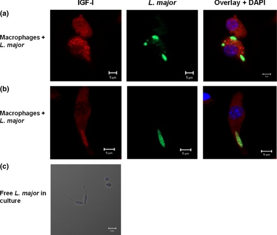
 ) with and without IFN-γ (200 U/mL) stimulus and with recombinant IGF-I (rIGF-I, 50 ng/mL) stimulus during a 48 h incubation. The data shown are representative of three independent assays. (d, e) Detection of IGF-I by confocal microscopy, using anti-IGF-I antibody (recognized by the secondary antibody AlexaFluor-546, shown in red) and anti-Leishmania antibody (recognized by the secondary antibody AlexaFluor-488, shown in green). 4′,6-diamidino-2-phenylindole (DAPI, shown in blue) was used to stain the nuclei. Images were captured using a confocal Leica LSM510 confocal microscope with a 63× objective and oil immersion. *P < 0·05 (
References
-
- Desjeux P. Leishmaniasis: current situation and new perspectives. Comp Immunol Microbiol Infect Dis. 2004;27:305–318. - PubMed
-
- Mougneau E, Bihl F, Glaichenhaus N. Cell biology and immunology of Leishmania. Immunol Rev. 2011;240:286–296. - PubMed
-
- Jones JI, Clemmons DR. Insulin-like growth factors and their binding proteins: biological actions. Endocr Rev. 1995;16:3–34. - PubMed
-
- Gomes CM, Goto H, Corbett CE, Gidlund M. Insulin-like growth factor-1 is a growth promoting factor for Leishmania promastigotes. Acta Trop. 1997;64:225–228. - PubMed
-
- Gomes CM, Monteiro HP, Gidlund M, Corbett CE, Goto H. Insulin-like growth factor-I induces phosphorylation in LeishmaniaLeishmaniamexicana promastigotes and amastigotes. J Eukaryot Microbiol. 1998;45:352–355. - PubMed
Publication types
MeSH terms
Substances
LinkOut - more resources
Full Text Sources
Other Literature Sources
