. 2013 Sep;173(3):512-22.
doi: 10.1111/cei.12138.

Disordered Toll-like receptor 2 responses in the pathogenesis of pulmonary sarcoidosis

Affiliations

Disordered Toll-like receptor 2 responses in the pathogenesis of pulmonary sarcoidosis

M I Gabrilovich et al. Clin Exp Immunol. 2013 Sep.

Abstract

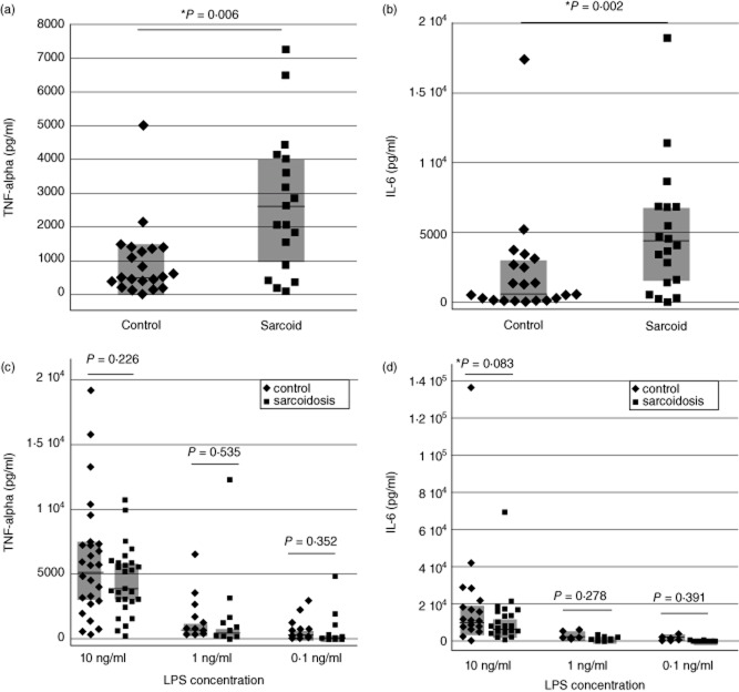
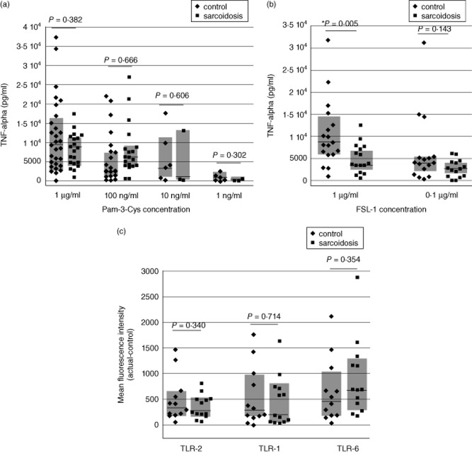
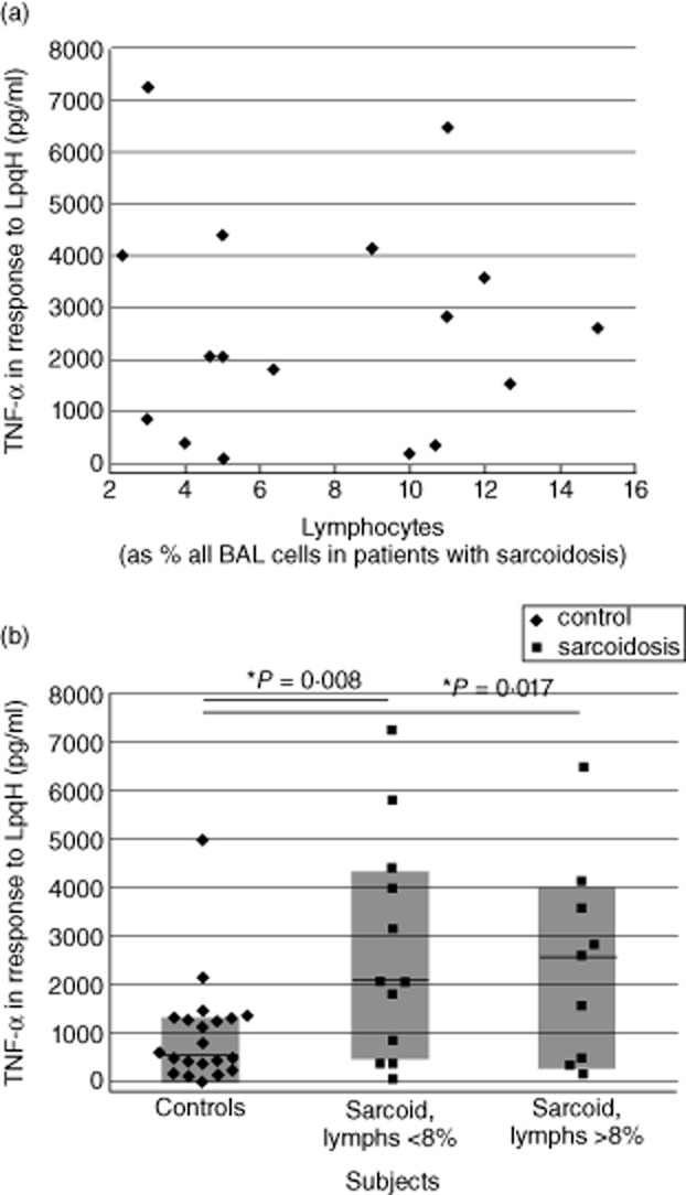
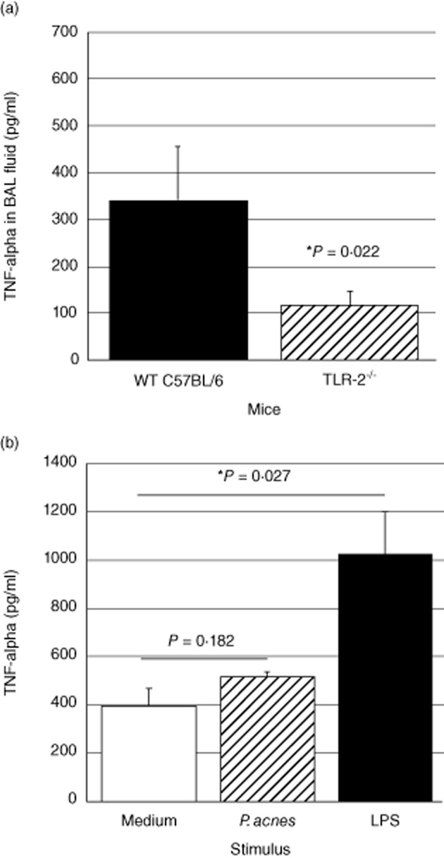
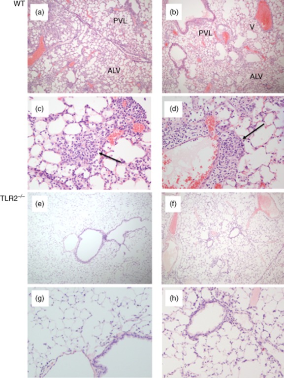
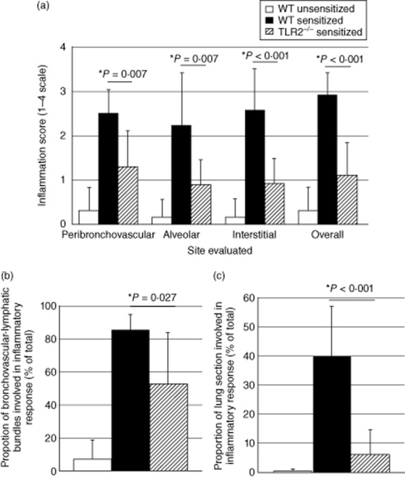
In this study, we hypothesized that the granulomatous disorder sarcoidosis is not caused by a single pathogen, but rather results from abnormal responses of Toll-like receptors (TLRs) to conserved bacterial elements. Unsorted bronchoalveolar lavage (BAL) cells from patients with suspected pulmonary sarcoidosis and healthy non-smoking control subjects were stimulated with representative ligands of TLR-2 (in both TLR-2/1 and TLR-2/6 heterodimers) and TLR-4. Responses were determined by assessing resulting production of tumour necrosis factor (TNF)-α and interleukin (IL)-6. BAL cells from patients in whom sarcoidosis was confirmed displayed increased cytokine responses to the TLR-2/1 ligand 19-kDa lipoprotein of Mycobacterium tuberculosis (LpqH) and decreased responses to the TLR-2/6 agonist fibroblast stimulating ligand-1 (FSL)-1. Subsequently, we evaluated the impact of TLR-2 gene deletion in a recently described murine model of T helper type 1 (Th1)-associated lung disease induced by heat-killed Propionibacterium acnes. As quantified by blinded scoring of lung pathology, P. acnes-induced granulomatous pulmonary inflammation was markedly attenuated in TLR-2(-/-) mice compared to wild-type C57BL/6 animals. The findings support a potential role for disordered TLR-2 responses in the pathogenesis of pulmonary sarcoidosis.

Keywords: Toll-like receptors; bronchoalveolar lavage (BAL); sarcoidosis.


Figures

Figure 1
Figure 1
Compared to those of healthy non-smokers, bronchoalveolar lavage (BAL) cells from sarcoidosis patients display greater cytokine responses to the Toll-like receptor (TLR)-2 ligand 19-kDa lipoprotein of Mycobacterium tuberculosis, but not to the TLR-4 ligand lipopolysaccharide (LPS). In response to stimulation the 19-kDa M. tuberculosis lipoprotein LpqH and the synthetic lipopeptide based on its structure (Pam-3-Cys-SSNKSTTGSGETTTA), peak levels of tumour necrosis factor (TNF)-α (a) and interleukin (IL)-6 (b) stimulation were both significantly greater for patients than control subjects. In contrast, no significant differences were observed between tumour necrosis factor (TNF)-α and IL-6 responses of BAL cells from sarcoidosis patients and from control subjects 24 h after stimulation with several concentrations of the TLR-4 ligand LPS, as detailed in the text (c,d, respectively). For all figures, horizontal lines indicate median values and shaded boxes indicate 25–75th percentile ranges.
Figure 2
Figure 2
Bronchoalveolar lavage (BAL) cells of sarcoidosis patients and healthy control subjects have differing responsiveness to stimulation of Toll-like receptor (TLR)-2 heterodimers, but do not display differential expression of TLR-2/1 and TLR-2/6. As detailed in the text, peak tumour necrosis factor (TNF)-α responses of BAL cells from patients and control subjects did not differ following stimulation with Pam-3-Cys (a). In contrast, BAL cells from sarcoidosis patients produced significantly less TNF-α than those of healthy controls in response to 1 μg/ml of the TLR-2/6 agonist fibroblast stimulating ligand-1 (FSL-1), but not in response to a lower concentration (0·1 μg/ml) of this ligand (b). Nevertheless, no differences were observed between expression of TLR-2, TLR-1 or TLR-6 on alveolar macrophages (AM) of control subjects and patients, as detailed in the text (c). For all figures, horizontal lines again indicate median results, whereas shaded boxes illustrate the 25–75th percentile range for each assessment.
Figure 3
Figure 3
Increased LpgH-induced production of tumour necrosis factor (TNF)-α by bronchoalveolar lavage (BAL) cells in sarcoidosis is independent of BAL cell lymphocytosis. As illustrated, BAL cell production of TNF-α shows no correlation with the percentage of lymphocytes present within BAL, (a). Further, division of these results into those observed for patients in whom lymphocytes accounted for greater or fewer than 8% of total BAL cells indicates that both groups display TNF-α responses that are distinct from those of control subjects (b).
Figure 4
Figure 4
Pulmonary tumour necrosis factor (TNF)-α responses to heat-killed Propionibacterium acnes are reduced in Toll-like receptor (TLR)-2–/– animals. Compared to similarly treated wild-type C57BL/6 animals, TLR-2–/– mice in which intratracheal (i.t.) P. acnes challenge was performed 14 days after initial intraperitoneal (i.p.) sensitization displayed significantly lower concentrations of TNF-α in bronchoalveolar lavage (BAL) fluid at 24 h. The data represent mean ± standard deviation (s.d.) of studies of six wild-type and seven TLR-2–/– animals (a). The specificity of this finding was clarified by in-vitro studies of lung homogenates from non-sensitized TLR-2–/– mice. As detailed in the text, stimulation with P. acnes did not induce significant TNF-α responses at 24 h, but stimulation with the TLR-4 ligand lipopolysaccharide (LPS) resulted in significant TNF-α production by these cells. Results represent mean ± s.d. of findings following incubation of three pools of homogenized lungs, each obtained from three TLR-2–/– mice (b).
Figure 5
Figure 5
Propionibacterium acnes-induced granulomatous pulmonary inflammation is reduced markedly in Toll-like receptor (TLR)-2–/– mice compared to that observed in wild-type C57BL/6 animals. Both groups of animals received intraperitoneal (i.p.) sensitization followed by intratracheal (i.t.) challenge with P. acnes. The images show representative histology of two wild-type animals at low (a,b) and high magnification (c,d). As illustrated, wild-type animals displayed a marked influx of lymphocytes and macrophages that were primarily adjacent to bronchovascular-lymphatic bundles (PVL) and large veins (V). Varying degrees of intra-alveolar and interstitial (ALV) inflammation were also observed. Magnified views show loosely formed granulomas typical of those observed in mice (indicated by arrows). In contrast, P. acnes induced relatively little inflammation in TLR-2–/– mice, as illustrated in (e) and (f) and the corresponding higher-power images (g) and (h). In these animals, peribronchovascular and lymphatic inflammation was minimal and intra-alveolar/interstitial regions were largely normal, as illustrated.
Figure 6
Figure 6
Detailed histological grading confirms marked reduction of pulmonary inflammation Toll-like receptor (TLR)-2 gene-disrupted animals following intratracheal (i.t.) challenge with Propionibacterium acnes. Pathology of non-sensitized and sensitized wild-type mice (n = 6 in each case) and of sensitized TLR-2–/– animals (n = 10) was evaluated. As detailed in the text, lungs of P. acnes-sensitized and challenged TLR-2–/– mice displayed significantly less inflammation than did sensitized, challenged wild-type mice in peribronchovascular, alveolar and interstitial regions. Overall inflammation scores were also significantly lower in TLR-2–/– mice (a). The percentage of peribronchovascular-lymphatic bundles and veins involved in the inflammatory response to P. acnes was significantly lower in TLR-2–/– mice than in sensitized wild-type animals (b). Similarly, the overall proportion of the lung tissue involved in the inflammatory response of TLR-2–/– mice was remarkably lower than that of wild-type mice and, in fact, did not differ significantly from that observed in non-sensitized wild-type mice (c). In all figures, findings in non-sensitized wild-type animals are represented by unshaded bars, sensitized wild-type animals by shaded bars and sensitized TLR-2–/– mice by striped bars.

Similar articles

Cited by

References

    1. Zissel G, Prasse A, Muller-Quernheim J. Immunologic response of sarcoidosis. Semin Respir Crit Care Med. 2010;321:390–403. - PubMed
    1. Saboor SA, Johnson NM, McFadden J. Detection of mycobacterial DNA in sarcoidosis and tuberculosis with polymerase chain reaction. Lancet. 1992;339:1012–1015. - PubMed
    1. Almenoff PL, Johnson A, Lesser M, Mattman LH. Growth of acid fast L forms from the blood of patients with sarcoidosis. Thorax. 1996;51:530–533. - PMC - PubMed
    1. Drake WP, Pei Z, Pride DT, Collins RD, Cover TL, Blaser MJ. Molecular analysis of sarcoidosis tissues for mycobacterium species DNA. Emerg Infect Dis. 2002;11:1334–1341. - PMC - PubMed
    1. Song Z, Marzilli L, Greenlee BM, et al. Mycobacterial catalase–peroxidase is a tissue antigen and target of the adaptive immune response in systemic sarcoidosis. J Exp Med. 2005;201:755–767. - PMC - PubMed

Publication types

MeSH terms