. 2013:3:1837.
doi: 10.1038/srep01837.

Microtubule alterations occur early in experimental parkinsonism and the microtubule stabilizer epothilone D is neuroprotective

Affiliations

Microtubule alterations occur early in experimental parkinsonism and the microtubule stabilizer epothilone D is neuroprotective

Daniele Cartelli et al. Sci Rep. 2013.

Abstract

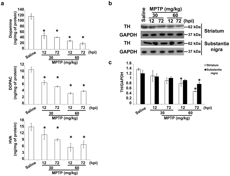
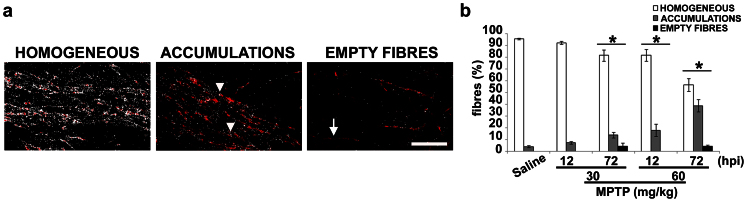
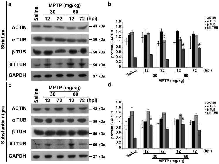
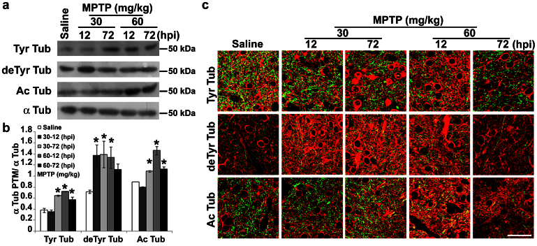
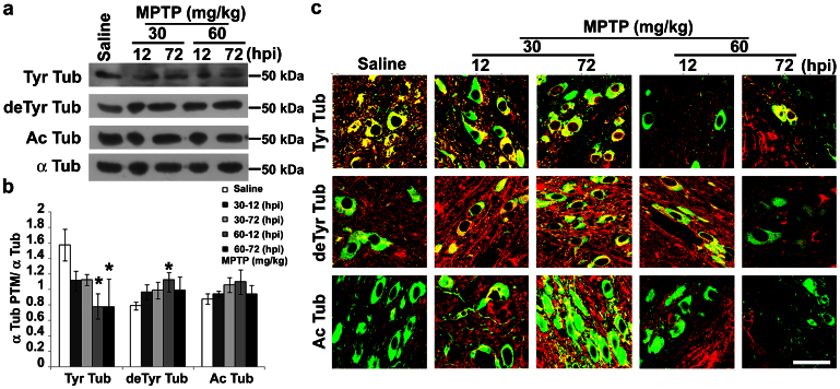
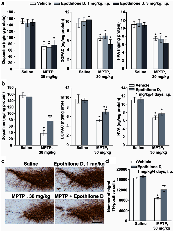
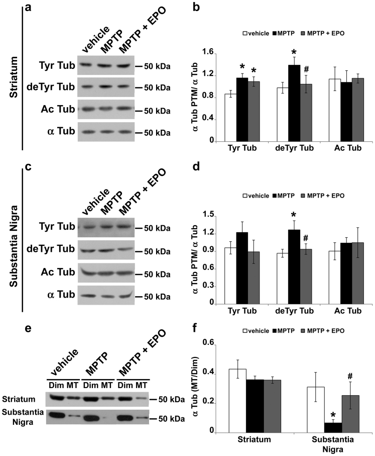
The role of microtubule (MT) dysfunction in Parkinson's disease is emerging. It is still unknown whether it is a cause or a consequence of neurodegeneration. Our objective was to assess whether alterations of MT stability precede or follow axonal transport impairment and neurite degeneration in experimental parkinsonism induced by 1-methyl-4-phenyl-1,2,3,6-tetrahydropyridine (MPTP) in C57Bl mice. MPTP induced a time- and dose-dependent increase in fibres with altered mitochondria distribution, and early changes in cytoskeletal proteins and MT stability. Indeed, we observed significant increases in neuron-specific βIII tubulin and enrichment of deTyr tubulin in dopaminergic neurons. Finally, we showed that repeated daily administrations of the MT stabilizer Epothilone D rescued MT defects and attenuated nigrostriatal degeneration induced by MPTP. These data suggest that alteration of ΜΤs is an early event specifically associated with dopaminergic neuron degeneration. Pharmacological stabilization of MTs may be a viable strategy for the management of parkinsonism.


Figures

Figure 1
Figure 1. Treatment paradigm underlines early alterations.
(a) Biochemical analyses of striatal dopamine, DOPAC and HVA levels in C57Bl mice injected with saline or MPTP (30 mg/kg, i.p., single injection or 20 mg/kg, i.p., × 3, 2 h apart) and killed 12 or 72 hours later (mean ± S.E.M., n = 8–10 mice per group). *P < 0.05; one-way ANOVA, Dunnett post hoc versus saline-injected mice. (b) Immunoblot of TH levels in lysates of the corpus striatum and substantia nigra of mice injected with saline or MPTP as in a. (c) Densitometric analyses of immunoblot reported in b (mean ± S.E.M., n = 8–10 mice per group). *P < 0.05; one-way ANOVA, Dunnett post hoc test versus saline-injected mice. hpi = hours post last injection of MPTP.
Figure 2
Figure 2. MPTP causes axonal transport impairment.
(a) Mask projections of sagittal sections of the nigrostriatal pathway in mice injected with saline or MPTP as in Fig. 1, showing the distribution of mitochondria (white spots) inside dopaminergic fibres (red). Arrowheads indicate mitochondria accumulations and arrow highlights an empty fibre. Scale bar = 20 μm. (b) Percentage of fibres displaying a homogeneous distribution of mitochondria, fibres showing dispersed or accumulated mitochondria or empty fibres. hpi = hours post last injection of MPTP (mean ± S.E.M., n = 9 sections from 3 different mice per group). *P < 0.05; χ2 test versus saline-injected mice.
Figure 3
Figure 3. MPTP treatment increases the βIII tubulin isotype.
(a) Immunoblots of actin, α-tubulin, β-tubulin and βIII tubulin in lysates of striatum of mice treated as in Fig. 1. (b) Densitometric analyses of immunoblot reported in a (mean ± S.E.M., n = 4–6 individuals per group). *P < 0.05; one-way ANOVA, Dunnett post hoc test versus saline-injected mice. (c) Immunoblot of actin, α-tubulin, β-tubulin and βIII tubulin in lysates of substantia nigra of mice treated as in Fig. 1. (d) Densitometric analyses of immunoblot reported in c (mean ± S.E.M., n = 4–6 individuals per group). *P < 0.05; one-way ANOVA, Dunnett post hoc test versus saline-injected mice. hpi = hours post last injection of MPTP.
Figure 4
Figure 4. MPTP affects MT stability in dopaminergic terminals.
(a) Immunoblot of levels of tyrosinated tubulin (Tyr Tub), detyrosinated tubulin (deTyr Tub) and acetylated tubulin (Ac Tub) in lysates of striatum of mice treated as in Fig. 1. (b) Densitometric analyses of immunoblot reported in a (mean ± S.E.M., n = 4–6 mice per group). For the quantitation, values of each α-tubulin PTM were normalized on the level of α-tubulin (α Tub) of the relative sample. *P < 0.05; one-way ANOVA, Dunnett post hoc test versus saline-injected mice. (c) Confocal images of striatum of mice treated as in Fig. 1. Green represents TH staining and red signals the various tubulin PTMs. Scale bar = 50 μm. hpi = hours post last injection of MPTP.
Figure 5
Figure 5. MPTP affects MT stability in dopaminergic neurons.
(a) Immunoblot of levels of tyrosinated tubulin (Tyr Tub), detyrosinated tubulin (deTyr Tub) and acetylated tubulin (Ac Tub) in lysates of substantia nigra of mice treated as in Fig. 1. (b) Densitometric analyses of immunoblot reported in a (mean ± S.E.M., n = 4–6 mice per group). For the quantitation, values of each α-tubulin PTM were normalized on the level of α-tubulin (α Tub) of the relative sample. *P < 0.05; one-way ANOVA, Dunnett post hoc test versus saline-injected mice. (c) confocal images of substantia nigra of mice treated as in Fig. 1. Green represents TH staining and red signals the various tubulin PTMs. Scale bar = 50 μm. hpi = hours post last injection of MPTP.
Figure 6
Figure 6. Repeated systemic injections of EpoD attenuate MPTP toxicity in mice.
(a) Biochemical analyses of striatal dopamine, DOPAC, and HVA levels in C57Bl mice injected with MPTP (30 mg/kg, i.p., single injection) alone or in combination with EpoD (1 or 3 mg/kg, i.p.) dissolved in DMSO and injected 30 min before MPTP. Mice were killed 7 days later (mean ± S.E.M., n = 8–10 mice per group). *P < 0.05; one-way ANOVA, Dunnett post hoc test versus saline-injected mice. (b) Biochemical analyses of striatal dopamine, DOPAC, and HVA levels in C57Bl mice injected with MPTP (30 mg/kg, i.p., single injection) alone or in combination with EpoD (1 mg/kg, i.p.) dissolved in DMSO and injected 30 min before MPTP and then for the following 4 days once a day. Mice were killed 7 days later (mean ± S.E.M., n = 8–10 mice per group). *, # P < 0.05; one-way ANOVA, Dunnett post hoc test versus saline-injected mice (*) or versus mice injected with MPTP alone (#). (c) Immunohistochemical analysis of TH in the pars compacta of substantia nigra of mice injected with a single i.p. injection of 30 mg/kg of MPTP, alone or combined with EpoD (1 mg/kg, i.p., 30 min prior to MPTP and then for the following 4 days, once a day). Scale bar = 250 μm. (d) Stereological counts of TH-positive cell in the substantia nigra pars compacta (mean ± S.E.M., n = 5 mice per group). *, # P < 0.05; one-way ANOVA, Dunnett post hoc test versus saline-injected mice (*) or versus mice injected with MPTP alone (#).
Figure 7
Figure 7. EpoD rescues MT system in MPTP-treated mice.
(a) Immunoblot of levels of tyrosinated tubulin (Tyr Tub), detyrosinated tubulin (deTyr Tub) and acetylated tubulin (Ac Tub) in lysates of striatum of mice treated as in Fig. 6. (b) Densitometric analyses of immunoblot reported in a (mean ± S.E.M., n = 5 mice per group). For the quantitation, values of each α-tubulin PTM were normalized on the level of α-tubulin (α Tub) of the relative sample. *, # P < 0.05; one-way ANOVA, Fischer LSD post hoc test versus saline-injected mice (*) or versus mice injected with MPTP alone (#). (c) Immunoblot of levels of tyrosinated tubulin (Tyr Tub), detyrosinated tubulin (deTyr Tub) and acetylated tubulin (Ac Tub) in lysates of substantia nigra of mice treated as in Fig. 6. (d) Densitometric analyses of immunoblot reported in c (mean ± S.E.M., n = 5 mice per group). For the quantitation, values of each α-tubulin PTM were normalized on the level of α-tubulin (α Tub) of the relative sample. *, # P < 0.05; one-way ANOVA, Fischer LSD post hoc test versus saline-injected mice (*) or versus mice injected with MPTP alone (#). Tubulin dimers (Dim) and MT polymers (MT) of corpus striatum and substantia nigra were analyzed by (e) immunoblot and (f) densitometric analyses and are shown as ratio (mean ± S.E.M., n = 3 mice per group). *, # P < 0.05; one-way ANOVA, Fischer LSD post hoc test versus saline-injected mice (*) or versus mice injected with MPTP alone (#).

References

    1. Sato M., Schwart W. H., Selden S. C. & Pollard T. D. Mechanical properties of brain tubulin and microtubules. J. Cell. Biol. 106, 1205–1211 (1988). - PMC - PubMed
    1. Mitchison T. & Kirschener M. Dynamic instability of microtubule growth. Nature 312, 237–242 (1984). - PubMed
    1. Inoué S. & Salmon E. D. Force generation by microtubule assembly/disassembly in mitosis and related movements. Mol. Biol. Cell 6, 1619–1640 (1995). - PMC - PubMed
    1. Conde C. & Caceres A. Microtubule assembly, organization and dynamics in axons and dendrites. Nat. Rev. Neurosci. 10, 319–332 (2009). - PubMed
    1. Baas P. W. & Black M. M. Individual microtubules in the axon consist of domain that differ both in composition and stability. J. Cell Biol. 111, 495–509 (1990). - PMC - PubMed

MeSH terms