

A novel vitreous substitute of using a foldable capsular vitreous body injected with polyvinylalcohol hydrogel
- PMID: 23670585
- PMCID: PMC3653144
- DOI: 10.1038/srep01838
A novel vitreous substitute of using a foldable capsular vitreous body injected with polyvinylalcohol hydrogel
Abstract
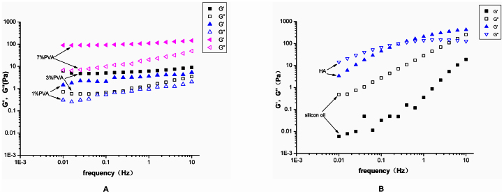
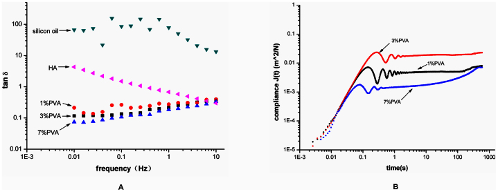
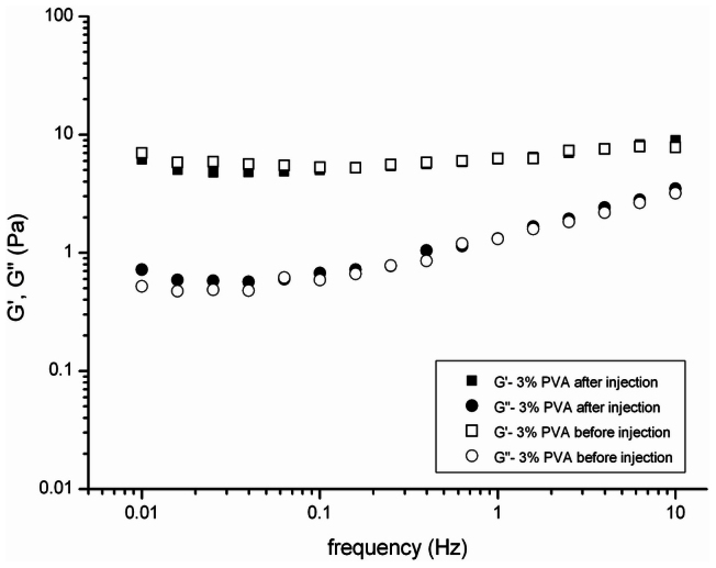
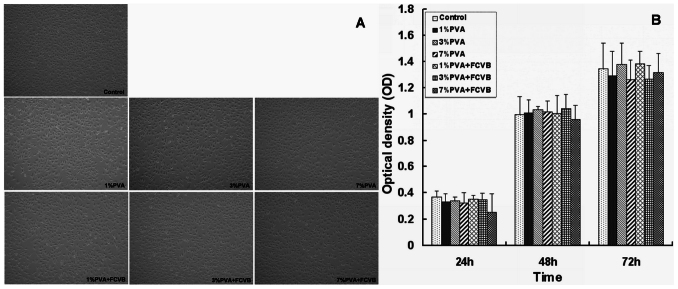
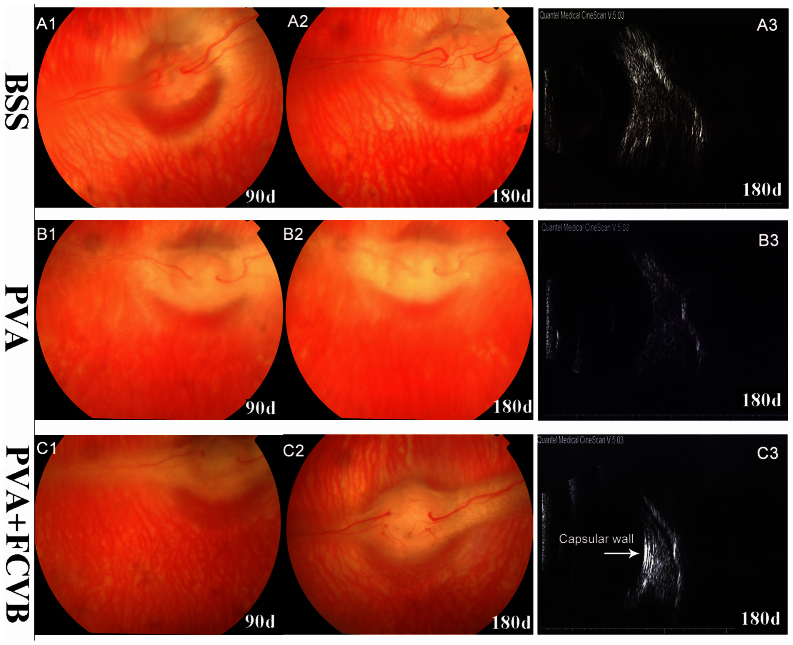
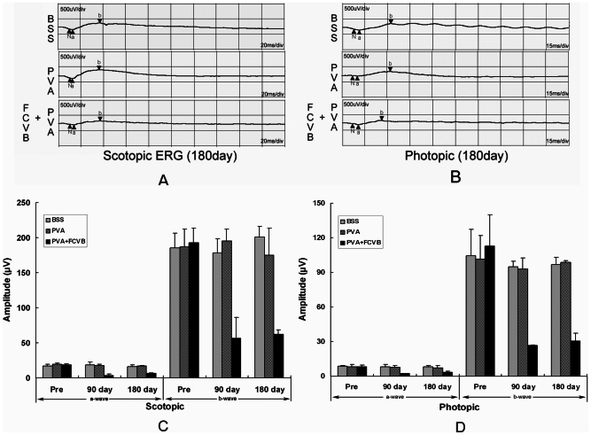
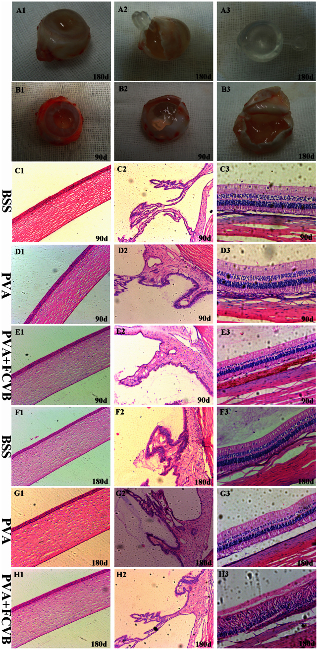
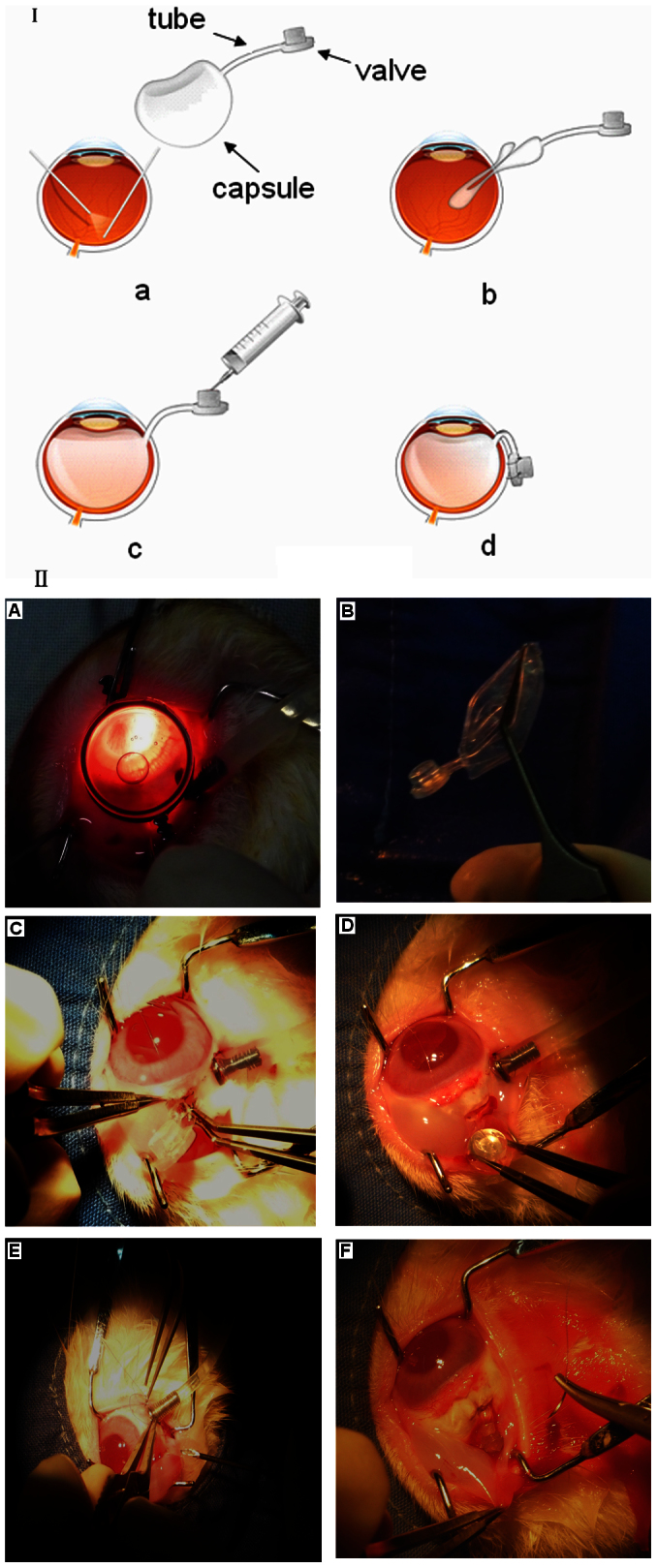
Hydrogels may be the ideal vitreous substitutes due to their wonderful physical features and biocompatibility. However, their drawbacks, short residence time, and biodegradation in vivo, have led to the fact that none of them have been approved for clinical use. In this study, we developed a novel approach of using a foldable capsular vitreous body (FCVB) injected with polyvinylalcohol (PVA) hydrogel as a vitreous substitute for long-term tamponade. The 3% PVA hydrogel that was cross-linked by gamma irradiation showed good rheological and physical properties and had no toxicity in vitro. After 180 days retention, the 3% PVA hydrogel inside FCVB remained transparent and showed good viscoelasticity without biodegradation and showed good biocompatibility and retina support. This new approach may develop into a valuable tool to improve the stability performance of PVA hydrogel as a vitreous substitute and to extend the application function of FCVB for long-term implantation in vitreous cavity.
Figures


Similar articles
-
Biocompatibility of polyvinylalcohol gel as a vitreous substitute.Curr Eye Res. 2006 Jul-Aug;31(7-8):599-606. doi: 10.1080/02713680600813854. Curr Eye Res. 2006. PMID: 16877268
-
Biocompatibility of polyvinyl alcohol/trisodium trimetaphosphate as vitreous substitute in experimental vitrectomy model in rabbits.J Biomed Mater Res B Appl Biomater. 2022 Feb;110(2):460-466. doi: 10.1002/jbm.b.34923. Epub 2021 Jul 29. J Biomed Mater Res B Appl Biomater. 2022. PMID: 34328263
-
Functional evaluation of a novel vitreous substitute using polyethylene glycol sols injected into a foldable capsular vitreous body.J Biomed Mater Res A. 2013 Sep;101(9):2538-47. doi: 10.1002/jbm.a.34560. Epub 2013 Jan 28. J Biomed Mater Res A. 2013. PMID: 23359564
-
Vitreous substitutes: An overview of the properties, importance, and development.J Biomed Mater Res B Appl Biomater. 2021 Aug;109(8):1156-1176. doi: 10.1002/jbm.b.34778. Epub 2020 Dec 14. J Biomed Mater Res B Appl Biomater. 2021. PMID: 33319466 Review.
-
Desired properties of polymeric hydrogel vitreous substitute.Biomed Pharmacother. 2024 Mar;172:116154. doi: 10.1016/j.biopha.2024.116154. Epub 2024 Feb 1. Biomed Pharmacother. 2024. PMID: 38306844 Review.
Cited by
-
Vitreous substitutes: challenges and directions.Int J Ophthalmol. 2015 Jun 18;8(3):437-40. doi: 10.3980/j.issn.2222-3959.2015.03.01. eCollection 2015. Int J Ophthalmol. 2015. PMID: 26085987 Free PMC article.
-
Vitreous Substitutes from Bench to the Operating Room in a Translational Approach: Review and Future Endeavors in Vitreoretinal Surgery.Int J Mol Sci. 2023 Feb 7;24(4):3342. doi: 10.3390/ijms24043342. Int J Mol Sci. 2023. PMID: 36834754 Free PMC article. Review.
-
Three-Year Efficacy and Safety of a Silicone Oil-Filled Foldable-Capsular-Vitreous-Body in Three Cases of Severe Retinal Detachment.Transl Vis Sci Technol. 2016 Feb 1;5(1):2. doi: 10.1167/tvst.5.1.2. eCollection 2016 Jan. Transl Vis Sci Technol. 2016. PMID: 26855843 Free PMC article.
-
The influence of UV-visible light, microwave radiation, argon laser, and heating and aging processes on silicone oil utilized as intravitreal implants: Experimental exposure with clinical correlation.PLoS One. 2024 Dec 31;19(12):e0316212. doi: 10.1371/journal.pone.0316212. eCollection 2024. PLoS One. 2024. PMID: 39739730 Free PMC article.
-
Self-Healing Injectable Hydrogels for Tissue Regeneration.Chem Rev. 2023 Jan 25;123(2):834-873. doi: 10.1021/acs.chemrev.2c00179. Epub 2022 Aug 5. Chem Rev. 2023. PMID: 35930422 Free PMC article. Review.
References
-
- Federman J. L. & Schubert H. D. Complications associated with the use of silicone oil in 150 eyes after retina-vitreous surgery. Ophthalmology. 95, 870–876 (1988). - PubMed
-
- Valone Jr J. & McCarthy M. Emulsified anterior chamber silicone oil and glaucoma. Ophthalmology. 101, 1908–1912 (1994). - PubMed
-
- Ichhpujani P., Jindal A. & Jay Katz L. Silicone oil induced glaucoma: a review. Graefes Arch Clin Exp Ophthalmol. 247, 1585–1593 (2009). - PubMed
Publication types
MeSH terms
Substances
LinkOut - more resources
Full Text Sources
Other Literature Sources
Medical
Miscellaneous
