. 2013 May 28;110(22):9130-5.
doi: 10.1073/pnas.1300454110. Epub 2013 May 13.

Perineuronal nets protect fast-spiking interneurons against oxidative stress

Affiliations

Perineuronal nets protect fast-spiking interneurons against oxidative stress

Jan-Harry Cabungcal et al. Proc Natl Acad Sci U S A. .

Abstract

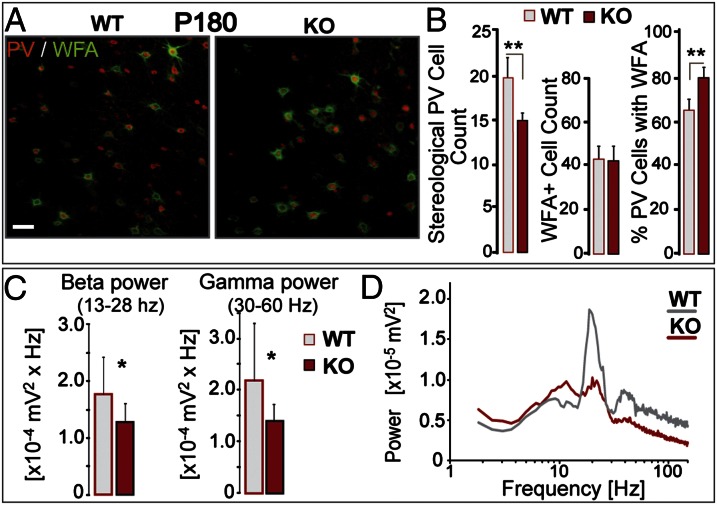
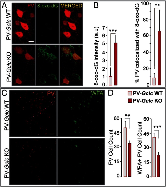
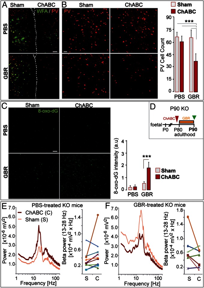
A hallmark of schizophrenia pathophysiology is the dysfunction of cortical inhibitory GABA neurons expressing parvalbumin, which are essential for coordinating neuronal synchrony during various sensory and cognitive tasks. The high metabolic requirements of these fast-spiking cells may render them susceptible to redox dysregulation and oxidative stress. Using mice carrying a genetic redox imbalance, we demonstrate that extracellular perineuronal nets, which constitute a specialized polyanionic matrix enwrapping most of these interneurons as they mature, play a critical role in the protection against oxidative stress. These nets limit the effect of genetically impaired antioxidant systems and/or excessive reactive oxygen species produced by severe environmental insults. We observe an inverse relationship between the robustness of the perineuronal nets around parvalbumin cells and the degree of intracellular oxidative stress they display. Enzymatic degradation of the perineuronal nets renders mature parvalbumin cells and fast rhythmic neuronal synchrony more susceptible to oxidative stress. In parallel, parvalbumin cells enwrapped with mature perineuronal nets are better protected than immature parvalbumin cells surrounded by less-condensed perineuronal nets. Although the perineuronal nets act as a protective shield, they are also themselves sensitive to excess oxidative stress. The protection might therefore reflect a balance between the oxidative burden on perineuronal net degradation and the capacity of the system to maintain the nets. Abnormal perineuronal nets, as observed in the postmortem patient brain, may thus underlie the vulnerability and functional impairment of pivotal inhibitory circuits in schizophrenia.

Keywords: critical period; extracellular matrix; glutamate cysteine ligase; glutathione; neuronal synchronization.


Conflict of interest statement

The authors declare no conflict of interest.

Figures

Fig. 1.
Fig. 1.
Chronic 6-mo redox dysregulation preferentially affects PV cells lacking PNNs. (A) PV immunoreactivity (red) and WFA-labeled PNNs (green) in the ACC of adult (P180) Gclm KO and WT mice. (Scale, 40 µm.) (B) (Left) stereological quantification reveals fewer PV immunoreactive cells in KO mice than in WT mice (n = 6 per group). (Center) WFA-labeled PNNs are not affected in KO mice. (Right) percentage of PV immunoreactive cells enwrapped by WFA-labeled PNNs is higher in KO mice, suggesting that the affected PV cells were those lacking PNNs. (C) Local neuronal synchronization in ACC slices (induced by carbachol, kainic acid, and quinpirole) is reduced in P180 KO mice. The power of β- (13–28 Hz; Left) and γ-oscillations (30–60 Hz; Right) is weaker in KO (n = 11) vs. WT slices (n = 15). (D) Power spectra of KO and WT recordings (mean of pooled data). Note that the peak frequencies of both β and γ oscillations do not differ significantly across genotypes [mean ± SD; β peak: 21.8 ± 3.2 Hz (KO) and 21.0 ± 2.3 Hz (WT); γ peak: 40.8 ± 5.6 Hz (KO) and 41.0 ± 4.5 Hz (WT)]. Bars, SD. **P < 0.01; *P < 0.05.
Fig. 2.
Fig. 2.
Mature PNNs protect PV cells from oxidative stress. (A) WFA-labeled PNNs in P20 Gclm KO mice are not fully developed around PV cells compared with P90 mice (n = 5 per group). Micrographs of PV immunoreactivity (red) and WFA labeling (green) in the ACC. (Scale, 40 µm.) (B) GBR-induced oxidative stress only affects PV cells in P20 Gclm KO. Micrographs of PV immunoreactivity in the ACC of P20 GBR- and PBS-treated KO mice. (Scale, 80 µm.) (C) GBR treatment generates significant additional oxidative stress in the ACC of P20 and P90 Gclm KO mice (n = 4 per group). Micrographs of 8-oxo-dG labeling after GBR treatment. (Scale, 40 µm.) (D) PV cells (red or arrowheads) surrounded by dense WFA-labeled PNNs (light blue, pseudocolor modified for improved visibility) display low levels of 8-oxo-dG signal (green), and vice versa. (Scale, 10 µm.) Quantile density contours and linear regression plot illustrate the inverse relationship between the number of voxels stained with WFA (PNNs) and 8-oxo-dG associated with each PV cell. Rank-transformed values for WFA (rWFA) and 8-oxo-dG (r8-oxo-dG) were used to compute the Pearson correlation coefficient. Bars, SD. **P < 0.01; *P < 0.05.
Fig. 3.
Fig. 3.
PNNs are sensitive to additional oxidative stress in Gclm KO mice. (A) Micrographs of PV (red) and WFA labeling (green) after GBR treatment in P90 Gclm KO and WT mice. (Scale, 40 µm.) (B) Quantification (n = 5 per group) shows that the number of PV immunoreactive cells is not significantly different in GBR-treated WT and KO mice (Left). However, GBR significantly reduces WFA labeling in KO compared with WT mice (Right). Bars, SD. ***P < 0.001.
Fig. 4.
Fig. 4.
PV cells and their PNNs are sensitive to cell-autonomous redox dysregulation. (A) Redox dysregulation restricted to PV cells (PV-Gclc KO mice) leads to strong 8-oxo-dG signals within PV cells of the ACC. Some cells without PV but with strong 8-oxo-dG labeling may have lost their PV immunoreactivity. (Scale, 10 μm.) (B) Quantification shows higher 8-oxo-dG labeling within the ACC (Left) and within PV cells (percentage PV colocalized with 8-oxo-dG; Right) of PV-Gclc KO compared with PV-Gclc WT mice (n = 5 per group). (C) PV immunoreactivity and WFA-labeled PNNs in the ACC of PV-Gclc KO and WT mice. (Scale, 50 μm.) (D) Quantification indicates significant decrease in number of PV immunoreactive cells (Left) and PNNs (Right) in PV-Gclc KO mice (n = 5 per group). Bars, SD. ***P < 0.001; **P < 0.01.
Fig. 5.
Fig. 5.
PNN removal by ChABC renders PV cells vulnerable to oxidative stress. (A) WFA (green) and PV (red) labeling after GBR or PBS treatment in ChABC-injected and contralateral sham ACC of P90 Gclm KO mice. Dashed line: separation of hemispheres (ChABC-injected ACC; Right). (Scale, 100 µm.) (B) GBR (n = 7), but not PBS (n = 5), induces a significant decrease in PV immunoreactive cells in ChABC-injected ACC. (Scale, 50 µm.) (C) The 8-oxo-dG labeling in ChABC-injected and contralateral sham ACC after GBR or PBS treatment. (Scale, 80 µm.) The 8-oxo-dG signal is stronger in ChABC-injected ACC after GBR treatment. (D) Experimental design. (E and F) Neuronal synchronization induced by coapplication of carbachol, kainic acid and quinpirole in PBS- (n = 5) (E) and GBR-treated (n = 5) (F) KO mice. The effect of PNN degradation on the oscillation power depends on the treatment (PBS vs. GBR): significant interaction (P = 0.039, multivariate analysis) between PNN status in ACC (ChABC- vs. sham-injected) and treatment (PBS vs. GBR). In PBS-treated mice, oscillations tend to be higher in ChABC-injected compared with contralateral sham ACC (9 of 11 slices) (E). In contrast, in GBR-treated mice, oscillations tend to be weaker in ChABC-injected compared with contralateral sham ACC (4 of 7 slices) (F). Graphs indicate the paired power values recorded in a ChABC-injected ACC and its contralateral sham ACC connected by a line. Bars, SD. ***P < 0.001.

References

    1. Sohal VS, Zhang F, Yizhar O, Deisseroth K. Parvalbumin neurons and gamma rhythms enhance cortical circuit performance. Nature. 2009;459(7247):698–702. - PMC - PubMed
    1. Whittington MA, Cunningham MO, LeBeau FE, Racca C, Traub RD. Multiple origins of the cortical γ rhythm. Dev Neurobiol. 2011;71(1):92–106. - PubMed
    1. Cabungcal JH, Steullet P, Kraftsik R, Cuenod M, Do KQ. Early-life insults impair parvalbumin interneurons via oxidative stress: Reversal by N-acetylcysteine. Biol Psychiatry. 2013;73(6):574–582. - PubMed
    1. Behrens MM, et al. Ketamine-induced loss of phenotype of fast-spiking interneurons is mediated by NADPH-oxidase. Science. 2007;318(5856):1645–1647. - PubMed
    1. Powell SB, Sejnowski TJ, Behrens MM. Behavioral and neurochemical consequences of cortical oxidative stress on parvalbumin-interneuron maturation in rodent models of schizophrenia. Neuropharmacology. 2012;62(3):1322–1331. - PMC - PubMed

Publication types

MeSH terms