. 2013 May 28;110(22):9118-23.
doi: 10.1073/pnas.1300003110. Epub 2013 May 13.

Palmitoylation-dependent CDKL5-PSD-95 interaction regulates synaptic targeting of CDKL5 and dendritic spine development

Affiliations

Palmitoylation-dependent CDKL5-PSD-95 interaction regulates synaptic targeting of CDKL5 and dendritic spine development

Yong-Chuan Zhu et al. Proc Natl Acad Sci U S A. .

Abstract

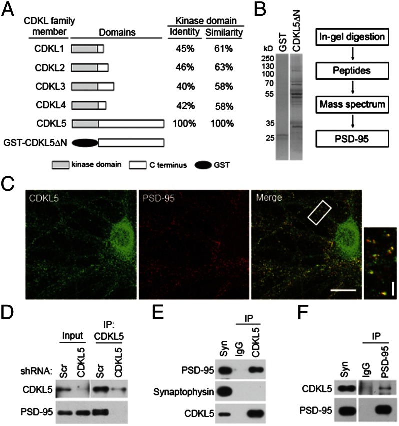
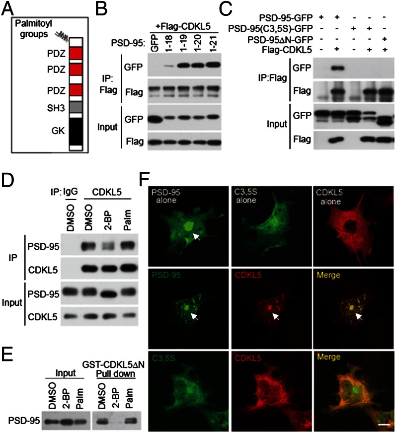
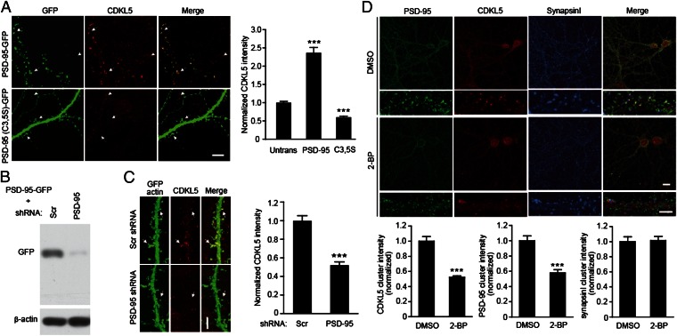
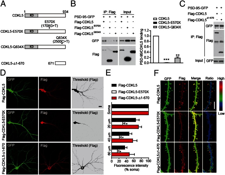
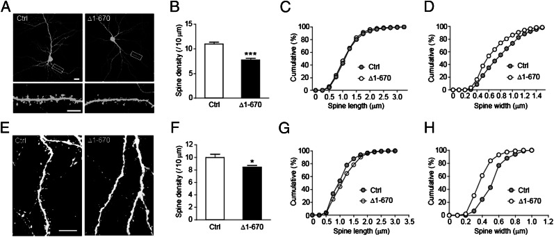
The X-linked gene cyclin-dependent kinase-like 5 (CDKL5) is mutated in severe neurodevelopmental disorders, including some forms of atypical Rett syndrome, but the function and regulation of CDKL5 protein in neurons remain to be elucidated. Here, we show that CDKL5 binds to the scaffolding protein postsynaptic density (PSD)-95, and that this binding promotes the targeting of CDKL5 to excitatory synapses. Interestingly, this binding is not constitutive, but governed by palmitate cycling on PSD-95. Furthermore, pathogenic mutations that truncate the C-terminal tail of CDKL5 diminish its binding to PSD-95 and synaptic accumulation. Importantly, down-regulation of CDKL5 by RNA interference (RNAi) or interference with the CDKL5-PSD-95 interaction inhibits dendritic spine formation and growth. These results demonstrate a critical role of the palmitoylation-dependent CDKL5-PSD-95 interaction in localizing CDKL5 to synapses for normal spine development and suggest that disruption of this interaction by pathogenic mutations may be implicated in the pathogenesis of CDKL5-related disorders.


Conflict of interest statement

The authors declare no conflict of interest.

Figures

Fig. 1.
Fig. 1.
PSD-95 is a CDKL5-interacting protein. (A) Schematic diagram for domain structures of CDKL family members and GST-CDKL5ΔN recombinant protein. (B) Isolation and identification of CDKL5-interacting proteins. (Left) Coomassie Bright Blue staining of SDS/PAGE gels for the purified proteins. (Right) Flow diagram of mass spectrum analysis. (C) Colocalization of CDKL5 and PSD-95 in hippocampal neurons. Neurons were fixed at 18 DIV and double-stained for CDKL5 and PSD-95. (Scale bar: 20 μm.) The boxed region is magnified to show the colocalization. (Scale bar: 5 μm.) (D) Interaction of CDKL5 and PSD-95 in neurons. Cultured neurons infected with lentivirus expressing scrambled (Scr) shRNA or CDKL5 shRNA were lysed and subjected to immunoprecipitation (IP) using a CDKL5 polyclonal antibody. The immunoprecipitates and the lysates were immunoblotted with CDKL5 or PSD-95 antibody. Interaction of CDKL5 and PSD-95 in the rat brain. Coimmunoprecipitations were performed in rat synaptosome (syn) using CDKL5 (E) or PSD-95 (F) antibody.
Fig. 2.
Fig. 2.
The CDKL5–PSD-95 interaction is controlled by palmitoylation. (A) Schematic diagram for domain structures of PSD-95. (B) Determination of the minimal binding region for CDKL5 in PSD-95. GFP or GFP-tagged PSD-95 derivatives (amino acids 1–18, 1–19, 1–20, and 1–21) were cotransfected with Flag-CDKL5 into 293T cells. Coimmunoprecipitation was performed in cell lysates with a Flag antibody. (C) Mutations preventing palmitoylation of PSD-95 abolish its interaction with CDKL5. PSD-95-GFP, PSD-95 (C3,5S)-GFP, and PSD-95ΔN-GFP were transfected individually or together with Flag-CDKL5 into 293T cells. Coimmunoprecipitation was performed in cell lysates with a Flag antibody. (D) Inhibition of palmitoylation decreases the interaction of CDKL5 with PSD-95 in neurons. Cortical neurons were treated with vehicle (DMSO), 20 μM 2-BP, or 20 μM palmitate for 8 h. After treatment, cells were harvested and the lysates were immunoprecipitated with a CDKL5 antibody. (E) GST pull-down experiment showing that the interaction of CDKL5 with PSD-95 is palmitoylation-dependent. Cortical neurons were treated as in D and the lysates were incubated with GST or GST-CDKL5ΔN affinity beads. Total lysates and precipitates were immunoblotted for PSD-95. (F) Palmitoylation-dependent interaction of CDKL5 and PSD-95 in COS-7 cells. COS-7 cells were transfected with PSD-95-GFP, PSD-95 (C3,5S)-GFP, and Flag-CDKL5, individually or together. Cells were fixed and stained for Flag. Arrows indicate the accumulation of proteins at the perinuclear region. (Scale bar: 20 μm.)
Fig. 3.
Fig. 3.
PSD-95 regulates synaptic targeting of CDKL5. (A) Effects of overexpression of WT or palmitoylation-deficient PSD-95 on synaptic localization of CDKL5. Hippocampal neurons were transfected with GFP-tagged WT PSD-95 or PSD-95 (C3,5S). Two days after transfection, cells were fixed and stained for CDKL5. (Scale bar: 5 μm.) Arrows and arrowheads indicate synaptic CDKL5 clusters in transfected and untransfected (Untrans) cells, respectively. Quantified fluorescence intensity data are shown in the bar graph (Right). A total of 20–30 dendritic segments from 10 to 15 neurons were quantified for each condition, ***P < 0.001, compared with untransfected cells. (B) Immunoblots showing the effectiveness of PSD-95 shRNA in down-regulating PSD-95-GFP expression in 293T cells. (C) Down-regulation of PSD-95 reduces synaptic CDKL5 levels. Arrows indicate synaptic CDKL5 clusters. (Scale bar: 5 μm.) n = 12–15 neurons for each condition; ***P < 0.001; t test. (D) Inhibition of palmitoylation reduces synaptic CDKL5 levels. Hippocampal neurons were treated with DMSO or 20 μM 2-BP for 8 h. Cells were fixed and stained for PSD-95, CDKL5, and synapsin I. n =15–20 microscope fields for each condition; ***P < 0.001, compared with DMSO treatment; t test.
Fig. 4.
Fig. 4.
Disease-associated mutations impair the interaction of CDKL5 with PSD-95 and its synaptic localization. (A) Schematic diagram showing the structures of CDKL5 and its mutants. KD, kinase domain. (B) Mutations E570× and Q834× impair the interaction of CDKL5 with PSD-95. The interaction was assayed by coimmunoprecipitation in 293T cells coexpressing GFP-tagged PSD-95 and Flag-tagged CDKL5 or its mutants. n = 3; **P < 0.01, ***P < 0.001. (C) Interaction of CDKL5 C-terminal region (CDKL5-Δ1-670) with PSD-95 assayed by coimmunoprecipitation. Subcellular localization of Flag-tagged CDKL5 and its mutants. (D) Representative images of neurons transfected with Flag-tagged CDKL5 or its mutants together with GFP, and stained for GFP and Flag. (Scale bar: 20 μm.) (E) Quantitation of average fluorescence in cell soma and in dendritic segments at the indicated distances from the soma in neurons expressing Flag-tagged proteins. n = 5–6 neurons for each condition; *P < 0.05 compared with Flag-CDKL5. (F) Representative images of dendrites showing the localization of Flag-tagged proteins in dendritic segments of transfected neurons. Ratio images indicate their enrichment in dendritic spines. (Scale bar: 5 μm.)
Fig. 5.
Fig. 5.
Disruption of CDKL5–PSD-95 interaction inhibits dendritic spine growth. (A) Representative images of hippocampal neurons transfected at DIV 8 with GFP together with empty vector or Flag-CDKL5-Δ1-670. Cells were fixed at DIV 15 and stained for GFP. [Scale bar: 20 μm (Top) and 5 μm (Bottom).] (B) Quantification of spine density in neurons transfected as indicated. n = 20–22 neurons for each; ***P < 0.001; t test. (C) Cumulative distribution of spine length plotted for the indicated conditions. P = 0.2586. Kolmogorov–Smirnov (K–S) test. (D) Cumulative distribution of spine width plotted for the indicated conditions. P < 0.001; K–S test. (E) Representative images of layer II–III pyramidal neurons in postnatal day (P) 14 rat brains transfected with GFP or GFP-CDKL5-Δ1-670 by in utero electroporation. Cells were stained with saturated GFP antibody to circumvent uneven distribution of GFP or GFP-tagged proteins in spines. (Scale bar: 10 μm.) (F) Quantification of spine density in neurons transfected as indicated. n = 5–6 neurons; *P < 0.05; t test. (G) Cumulative distribution of spine length plotted for the indicated conditions. P = 0.0414; K–S test. (H) Cumulative distribution of spine width plotted for the indicated conditions. P < 0.001; K–S test.

References

    1. Scala E, et al. CDKL5/STK9 is mutated in Rett syndrome variant with infantile spasms. J Med Genet. 2005;42(2):103–107. - PMC - PubMed
    1. Tao J, et al. Mutations in the X-linked cyclin-dependent kinase-like 5 (CDKL5/STK9) gene are associated with severe neurodevelopmental retardation. Am J Hum Genet. 2004;75(6):1149–1154. - PMC - PubMed
    1. Malumbres M, et al. Cyclin-dependent kinases: A family portrait. Nat Cell Biol. 2009;11(11):1275–1276. - PMC - PubMed
    1. Montini E, et al. Identification and characterization of a novel serine-threonine kinase gene from the Xp22 region. Genomics. 1998;51(3):427–433. - PubMed
    1. Chen Q, et al. CDKL5, a protein associated with rett syndrome, regulates neuronal morphogenesis via Rac1 signaling. J Neurosci. 2010;30(38):12777–12786. - PMC - PubMed

Publication types

MeSH terms

Substances

LinkOut - more resources