. 2013 Jun 18;110(25):10252-7.
doi: 10.1073/pnas.1301480110. Epub 2013 May 13.

Dominant suppression of inflammation by glycan-hydrolyzed IgG

Affiliations

Dominant suppression of inflammation by glycan-hydrolyzed IgG

Kutty Selva Nandakumar et al. Proc Natl Acad Sci U S A. .

Retraction in

Abstract

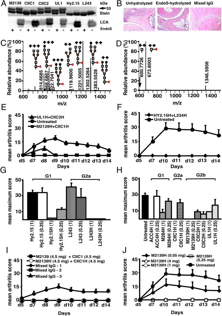
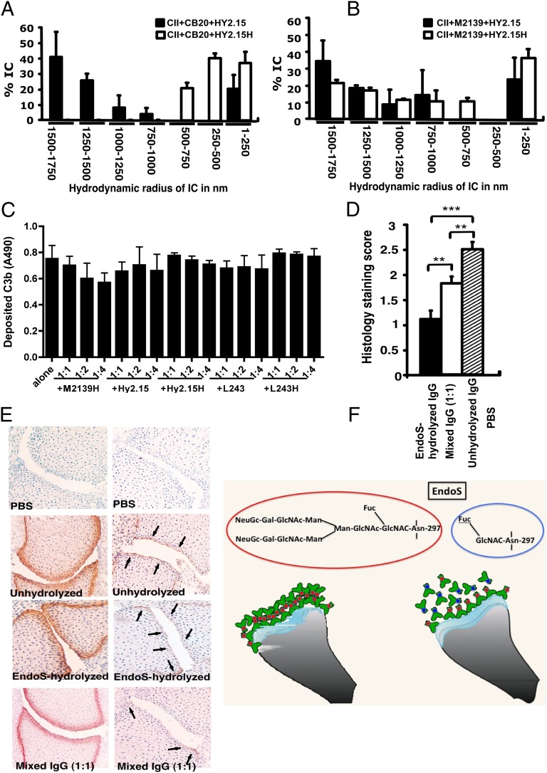
A unique anti-inflammatory property of IgG, independent of antigen specificity, is described. IgG with modification of the heavy-chain glycan on asparagine 297 by the streptococcal enzyme endo-β-N-acetylglucosaminidase (EndoS) induced a dominant suppression of immune complex (IC)-mediated inflammation, such as arthritis, through destabilization of local ICs by fragment crystallizable-fragment crystallizable (Fc-Fc) interactions. Small amounts (250 µg) of EndoS-hydrolyzed IgG were sufficient to inhibit arthritis in mice and most effective during the formation of ICs in the target tissue. The presence of EndoS-hydrolyzed IgG disrupted larger IC lattice formation both in vitro and in vivo, as visualized with anti-C3b staining. Neither complement binding in vitro nor antigen-antibody binding per se was affected.

Keywords: collagen; endoglycosidase; glycosylation; monoclonal antibody; rheumatoid arthritis.


Conflict of interest statement

Conflict of interest statement: Hansa Medical AB has filed patent applications for using EndoS-modified IgG for treatment of arthritis and K.S.N., M.C., and R.H. are listed as inventors. The authors have no additional financial interests. The funders had no role in the preparation of the manuscript or the decision to publish.

Figures

Fig. 1.
Fig. 1.
EndoS-hydrolyzed IgG dominantly inhibit inflammation. (A) SDS/PAGE and lectin blot analysis of mAbs incubated with (+) or without (−) EndoS hydrolysis and separated by 10% SDS/PAGE. The proteins were detected by PageBlue stain (Stain) or by blotting onto a PVDF membrane probed with Lens culinaris agglutinin (LCA). (B) Representative figures of H&E-stained ankle joints of mice (n = 3–4 per group) injected with anti-CII mAbs; unhydrolyzed (Left), EndoS-hydrolyzed (Center), or mixed IgG (Right). Magnification ×10. (C) Hy2.15 and (D) EndoS-treated Hy2.15. Shown spectra were acquired during the time period for which the majority of glycosylated peptides from EEQFNSTFR (21.5–23.0 min) elute. Doubly and triply charged ions as well as predicted glycan structures are shown. All numbers given are for the monoisotopic mass charge. In all of the animal experiments, male (BALB/c × B10.Q) F1 mice were used. Unless otherwise stated all of the mice received antibodies i.v. (d 0) and 25 μg of LPS i.p. (d 5). For arthritis induction in experiments shown in E, F, I and J, 9 mg of two anti-CII mAb mixtures (M2139 + CIIC1) were used, whereas for experiments in G and H 4 mg of four anti-CII mAb mixture (M2139 + CIIC1 + CIIC2 + UL1) was used. Antigen specificity is not required for inhibition. Mice (n = 42) were injected with 4 mg of EndoS-hydrolyzed IgG (E) M2139H + CIIC1H or UL1H + CIIC2H or (F) Hy2.15H + L243H followed by anti-CII mAb. Dose and subclass dependency. (G) Mice (n = 39) were injected with EndoS-hydrolyzed or unhydrolyzed IgG1 (Hy2.15) or IgG2a (L243) mAb binding to joint unrelated antigens at two different concentrations (1 mg and 0.25 mg), followed by anti-CII mAb. (H) Mice (n = 65) were injected with different subclasses of EndoS-hydrolyzed anti-CII (M284H, M2139H, CIIC1H, CIIC2H, and UL1H) or anti-citrullinated CII peptide IgG (ACC4H) at two different concentrations (1 mg and 0.25 mg), followed by anti-CII mAb. (I) Mice (n = 25) were injected with a mixture of EndoS-hydrolyzed and/or unhydrolyzed anti-CII IgG at different combinations. In mixed IgG groups, group 1 received 4.5 mg of unhydrolyzed and 4.5 mg of EndoS-hydrolyzed IgG, group 2 had 6.75 mg of unhydrolyzed and 2.25 mg of EndoS-hydrolyzed IgG, and group 3 received 7.8775 mg of unhydrolyzed and 1.125 mg of EndoS-hydrolyzed IgG. (J) Mice (n = 25) were injected with different concentrations (50–4,000 μg) of EndoS-hydrolyzed single anti-CII IgG (M2139H), followed by anti-CII mAb. Three hours after the antibody transfer, LPS was injected. H denotes EndoS-hydrolyzed IgG. Hy2.15 and L243 represent mAbs binding to TNP hapten and human HLA-DR antigen, respectively. Error bars indicate ± SEM.
Fig. 2.
Fig. 2.
Inhibition of inflammation and SPR and FTIRM analysis. (A) Mice (n = 30) were injected with 1 mg of EndoS-hydrolyzed anti-CII IgG (M2139H + CIIC1H +CIIC2H + UL1H) at different time points (−48, −3, 0, +3, or +48 h). At 0 h and 3 h, anti-CII mAb (M2139 + CIIC1 + CIIC2 + UL1) and then LPS were injected. One group of mice received no treatment. (B) Effect of splenectomy. Mice (n = 21) were either splenectomized (Splx) or sham-operated (Sham). Three weeks later, they were injected with 4 mg of EndoS-hydrolyzed IgG (M2139H + CIICH) or left untreated, followed by anti-CII mAb (M2139 + CIIC1). H denotes EndoS-hydrolyzed IgG. Error bars indicate ±SEM. (C) SPR (Biacore) analysis of antibody binding capacity of EndoS-hydrolyzed and unhydrolyzed IgG was performed using CII immobilized on CM5 sensor chip. MAbs were injected at different concentrations through flow cells at a flow rate of 30 μL/min. Antibodies were injected for 3 min and dissociation of bound molecules was observed for 7 min. There was no difference in antibody binding when EndoS-hydrolyzed or unhydrolyzed IgGs were added at different ratios to anti-CII mAb mixture. (D and E) Changes in the chemical composition of the cartilage were assessed using FTIRM analysis. Representative mean spectra are shown from cartilage cultures without antibody (D), and from cartilage cultured for 14 d with 100 μg/mL of unhydrolyzed mAb M2139 (E). The results shown are the mean of 10 measurements taken from the central areas (red line) and near the surface of the tissue (black line). The mean spectra for surface and interior were calculated to assess the effects of antibody penetration on the peaks characteristic of CII and of proteoglycans. (F and G) The mean peaks from the surface cartilage were compared with those from antibody-exposed surface of cartilage exposed to the EndoS-hydrolyzed or unhydrolyzed IgG. Cartilage exposed to either EndoS-hydrolyzed or unhydrolyzed IgG (CIIC1, M2139, and UL1) showed similar changes. (F) The height and location of the amide 1 peak, which represents the total protein content of the tissue, in the region 1,600–1,700 cm–1. (G) The height of the peak at 1,076 cm−1 represents proteoglycans.
Fig. 3.
Fig. 3.
Disturbance of stable ICs and complement activation. CII (1 mg/mL) and anti-CII mAb CB20 (low affinity, A) or (M2139 (high affinity, B) at 1 mg/mL were mixed together at 1:1 ratio and incubated for 30 min at 37 °C, followed by addition of anti-hapten IgG, either unhydrolyzed (Hy2.15) or EndoS-hydrolyzed IgG (Hy2.15H) at the ratio 1:1:1. Twenty microliters of this mixture were loaded onto the capillary tube in the DLS instrument. Relative sizes of ICs present in the solution are indicated in the x-axis and the y-axis denotes percentage of ICs present in the solution. Each sample was measured five to seven times and the bars represent mean values from two experiments. Error bars indicate ± SEM. (C) Complement activation on CII bound anti-CII antibodies were monitored by measuring C3b deposition. Each bar represents mean values from three experiments ± SD. (D) Deposition of C3b on the cartilage of mice was used as a measure of IC deposition and complement activation after the injection of EndoS-hydrolyzed, unhydrolyzed, or mixed anti-CII IgG. Mouse pups (three or four mice per group) were injected with 1 mg each of unhydrolyzed IgG (M2139 + CIIC2 + UL1), EndoS-hydrolyzed IgG (M2139H + CIIC2H + UL1H), or a mixture of IgGs at 1:1 ratio. In each group, 26–44 joints were scored in total. **P < 0.01; ***P < 0.005. Error bars indicate ± SEM. (E) Paw samples collected 24 h later were stained with biotinylated anti-kappa (Left) or goat anti-mouse anti-C3c antibodies (Right). Joint sections from mice injected with PBS (first row), unhydrolyzed (second row), EndoS-hydrolyzed (third row), or a mixture (1:1) of IgG (fourth row) are shown. Magnification ×20. Arrows indicate C3b deposition within ICs formed on the joint cartilage surface. (F) Diagram illustrating possible binding mechanisms involved in the suppression of arthritis by EndoS-hydrolyzed antibodies.

Comment in

  • Therapeutic potential of deglycosylated antibodies.
    Crispin M. Crispin M. Proc Natl Acad Sci U S A. 2013 Jun 18;110(25):10059-60. doi: 10.1073/pnas.1307776110. Epub 2013 Jun 7. Proc Natl Acad Sci U S A. 2013. PMID: 23749872 Free PMC article. No abstract available.

Similar articles

Cited by

References

    1. Ferrara C, et al. Unique carbohydrate-carbohydrate interactions are required for high affinity binding between FcgammaRIII and antibodies lacking core fucose. Proc Natl Acad Sci USA. 2011;108(31):12669–12674. - PMC - PubMed
    1. Deisenhofer J. Crystallographic refinement and atomic models of a human Fc fragment and its complex with fragment B of protein A from Staphylococcus aureus at 2.9- and 2.8-A resolution. Biochemistry. 1981;20(9):2361–2370. - PubMed
    1. Barb AW, Prestegard JH. NMR analysis demonstrates immunoglobulin G N-glycans are accessible and dynamic. Nat Chem Biol. 2011;7(3):147–153. - PMC - PubMed
    1. Parekh RB, et al. Association of rheumatoid arthritis and primary osteoarthritis with changes in the glycosylation pattern of total serum IgG. Nature. 1985;316(6027):452–457. - PubMed
    1. Kaneko Y, Nimmerjahn F, Ravetch JV. Anti-inflammatory activity of immunoglobulin G resulting from Fc sialylation. Science. 2006;313(5787):670–673. - PubMed

Publication types

MeSH terms