. 2013 May 28;110(22):E2028-37.
doi: 10.1073/pnas.1219956110. Epub 2013 May 13.

Molecular evolution of peptidergic signaling systems in bilaterians

Affiliations

Molecular evolution of peptidergic signaling systems in bilaterians

Olivier Mirabeau et al. Proc Natl Acad Sci U S A. .

Abstract

Peptide hormones and their receptors are widespread in metazoans, but the knowledge we have of their evolutionary relationships remains unclear. Recently, accumulating genome sequences from many different species have offered the opportunity to reassess the relationships between protostomian and deuterostomian peptidergic systems (PSs). Here we used sequences of all human rhodopsin and secretin-type G protein-coupled receptors as bait to retrieve potential homologs in the genomes of 15 bilaterian species, including nonchordate deuterostomian and lophotrochozoan species. Our phylogenetic analysis of these receptors revealed 29 well-supported subtrees containing mixed sets of protostomian and deuterostomian sequences. This indicated that many vertebrate and arthropod PSs that were previously thought to be phyla specific are in fact of bilaterian origin. By screening sequence databases for potential peptides, we then reconstructed entire bilaterian peptide families and showed that protostomian and deuterostomian peptides that are ligands of orthologous receptors displayed some similarity at the level of their primary sequence, suggesting an ancient coevolution between peptide and receptor genes. In addition to shedding light on the function of human G protein-coupled receptor PSs, this work presents orthology markers to study ancestral neuron types that were probably present in the last common bilaterian ancestor.

Keywords: GPCR evolution; bilaterian CNS cell types; bilaterian brain evolution; neuropeptide evolution.


Conflict of interest statement

The authors declare no conflict of interest.

Figures

Fig. 1.
Fig. 1.
Phylogenomics pipeline for the study of PS evolution. A standard phylogenomics strategy was used to derive sets of potential peptide GPCRs (receptor search, A) for all of the species considered (Methods). The final three phylogenetic trees of bilaterian rhodopsin GPCRs (β and γ rGPCRs and sGPCRs) were used to derive potential ancestral PSs. Then, to isolate potential peptide precursor sequences (peptide search, B) a noncanonical strategy was used, which involved the use of an HMM designed to find candidate peptide precursors in each of the bilaterian species and the construction of peptide phylogenetic trees using the neighbor-joining method and a normalized kernel-based distance (Methods).
Fig. 2.
Fig. 2.
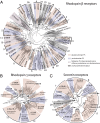
Phylogenetic analysis of bilaterian rhodopsin and secretin receptors. Maximum likelihood tree of bilaterian rhodopsin β (A), γ-type (B), and secretin (C) receptors, according to the GRAFS classification established in ref. . The tree is structured in well-supported subtrees containing both clusters of protostome (blue) and deuterostome (pink) groups of sequences. At the root of blue-pink subtrees (shown as black or green solid circles), a prototypic receptor of each subtype was already present in the urbilaterian. Black solid circles indicate well-supported bilaterian GPCR families, and green solid circles show hypothetical evolutionary relationships among bilaterian families. The bilaterian (b-), protostomian (p-), deuterostomian (d-), chordate (c-), lophotrochozoan (-l), or arthropod (a-) origin is indicated by an initial letter before each peptide GPCR acronym. Ancestral bilaterian clusters containing receptors characterized only in either protostomes or deuterostomes (e.g., b-TRHR and b-ETHR) were colored with alternating blue and pink bands, and bilaterian clusters containing no characterized receptors were shaded in gray. Photoreceptors and aminergic receptors were used as an outgroup for rhodopsin β receptors (A), and human adhesion GPCRs were used as an outgroup for the secretin receptors (C).
Fig. 3.
Fig. 3.
Conserved introns in β-rhodopsin receptor genes. Motif logo of rhodopsin β receptor alignment showing the introns that have a conserved position across bilaterians. Names of deuterostome, protostome, or bilaterian PSs were used, as defined in Fig. 2. The seven transmembrane domains (TM1–7) are indicated by dashed boxes. Single, double, and triple arrows indicate that the intron phase is 0, 1, or 2, respectively.
Fig. 4.
Fig. 4.
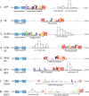
Conservation of eight ancestral bilaterian peptide precursor families. Conserved features of these eight peptide precursor families include key residues shown in the peptide logo. For example, the N-terminal glutamine in GnRH/AKH sequences, pairs of cysteines in AVP and Calc/DH31 sequences, the position of peptide(s) and other domains inside the precursor sequence, and constrained length distribution of spacer sequences in the precursor (shown by histograms).
Fig. 5.
Fig. 5.
Evolutionary scenario for the Ox and AT precursors. Structure of the hypothetical bilaterian ancestral Ox/AT precursor. (A) The hypothetical ancestral Ox/AT bilaterian precursor is composed of an N-terminal signal peptide (blue box), an Ox or AT peptide, represented by the two logo motifs just C-terminal to the signal peptide, and a C-terminal domain of unknown function that is found in most protostomes and in a deuterotostome, the acorn worm. However, we cannot conclude whether this precursor was more closely related to the extant deuterostome Ox neuropeptides bearing prototypic cysteine patterns or to the extant protostomian AT neuropeptides. (B) Probable scenario describing Ox/AT precursor evolution. Even though Oxs (red half-circle) and ATs (yellow half-circle) display no obvious similarity, their receptors are orthologous to each other and the last common ancestor of bilaterians possessed a C-terminal domain (orange triangle) that was retained in present-day ambulacrarians and protostomes and was lost in the lineage leading to chordates.
Fig. 6.
Fig. 6.
Ancestral bilaterian peptidergic systems. Inferred evolutionary relationships between the different ancestral bilaterian PSs. (A) Names of characterized deuterostomian systems (Left) with their orthologous protostomian systems (Right). A top-left half-square indicates the presence of the peptide in a given phylogenetic group, or single species, in the case of Branchiostoma (floridae) and Daphnia (pulex). Subphylum Vertebrata is composed of H. sapiens and Takifugu rubripes, phylum Tunicata of Ciona intestinalis and Ciona savignyi, superphylum Ambulacraria of S. purpuratus and S. kowalevskii, Lophotrochozoa of C. teleta and L. gigantea, class Insecta of D. melanogaster, Tribolium castaneum and Acyrthosiphon pisum and phylum Nematoda of C. elegans and Pristionchus pacificus. A bottom-right half-square indicates the presence of the receptor in a group. A receptor was considered to be present in a given group of animals when it was positioned inside a well-supported subtree (branch support value >0.95) including at least one characterized receptor. A full square denotes the presence of both peptides and receptors from a given PS. A green full square indicates that, for at least one member of that group, binding between a peptide and its receptor was biochemically demonstrated. For both peptides and receptors, the presence in a phylogenetic group implied most of the time the presence of at least two members of that group clustered together in the peptidergic system-specific subtree. The plus symbol refers to the last common ancestor of the given systems and a slash indicates the different names that were given to orthologous systems in distinct species. (B) Branch support values for each of the subclasses of peptide receptors are represented as solid circles of area scaled according to the three statistical test values: likelihood ratio test (PValSH) and bootstrap (btspML) values for maximum likelihood trees and posterior probability values (PPBayes) for the Bayesian reconstruction trees. (C) For each bilaterian PS, we wrote down the positions (relative to the global alignment of rhodopsin β receptors) and phases of introns that were conserved in, and specific to, that PS (Fig. 3). (D) For each peptide family we reported the largest phylogenetic group level (A, arthropods; B, bilaterians; C, chordates; D, deuterostomes; P, protostomes) for which peptide or precursor similarity could be detected in alignments (see also Dataset S1). An asterisk after the letter indicates the presence of a conserved domain outside of the peptide region that was used to establish our orthology hypotheses. Notable phyla-specific losses, expansions, and appearances of known PS: (1) AVP was lost in Drosophila. (2) AT was lost in Drosophila. (3) NPS was lost in teleosts. (4) A large expansion of both NPFF peptide and receptor genes is observed in amphioxus. (5) Large expansion of both Kiss1 peptide and receptor genes in amphioxus. (6) PTH-like peptides and glucagon-like peptides are found in Ciona and Branchiostoma.( 7) Receptors from the PTH + glucagon + PACAP family are absent from the genome of Drosophila.

Similar articles

Cited by

References

    1. Bockaert J, Pin JP. Molecular tinkering of G protein-coupled receptors: An evolutionary success. EMBO J. 1999;18(7):1723–1729. - PMC - PubMed
    1. Krishnan A, Almén MS, Fredriksson R, Schiöth HB. The origin of GPCRs: Identification of mammalian like Rhodopsin, Adhesion, Glutamate and Frizzled GPCRs in fungi. PLoS ONE. 2012;7(1):e29817. - PMC - PubMed
    1. Fredriksson R, Lagerström MC, Lundin LG, Schiöth HB. The G-protein-coupled receptors in the human genome form five main families. Phylogenetic analysis, paralogon groups, and fingerprints. Mol Pharmacol. 2003;63(6):1256–1272. - PubMed
    1. Douglass J, Civelli O, Herbert E. Polyprotein gene expression: Generation of diversity of neuroendocrine peptides. Annu Rev Biochem. 1984;53:665–715. - PubMed
    1. Steiner DF. The proprotein convertases. Curr Opin Chem Biol. 1998;2(1):31–39. - PubMed

Publication types

LinkOut - more resources