. 2013 Jul;33(14):2718-31.
doi: 10.1128/MCB.01254-12. Epub 2013 May 13.

Tumor necrosis factor α-induced hypoxia-inducible factor 1α-β-catenin axis regulates major histocompatibility complex class I gene activation through chromatin remodeling

Affiliations

Tumor necrosis factor α-induced hypoxia-inducible factor 1α-β-catenin axis regulates major histocompatibility complex class I gene activation through chromatin remodeling

Sadashib Ghosh et al. Mol Cell Biol. 2013 Jul.

Abstract

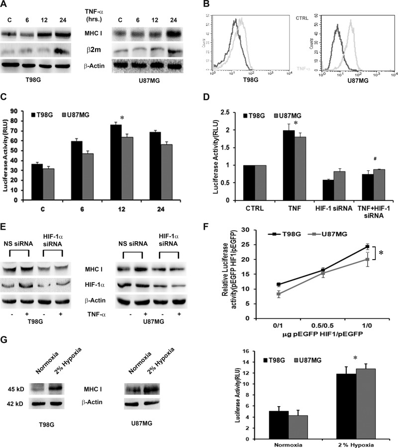
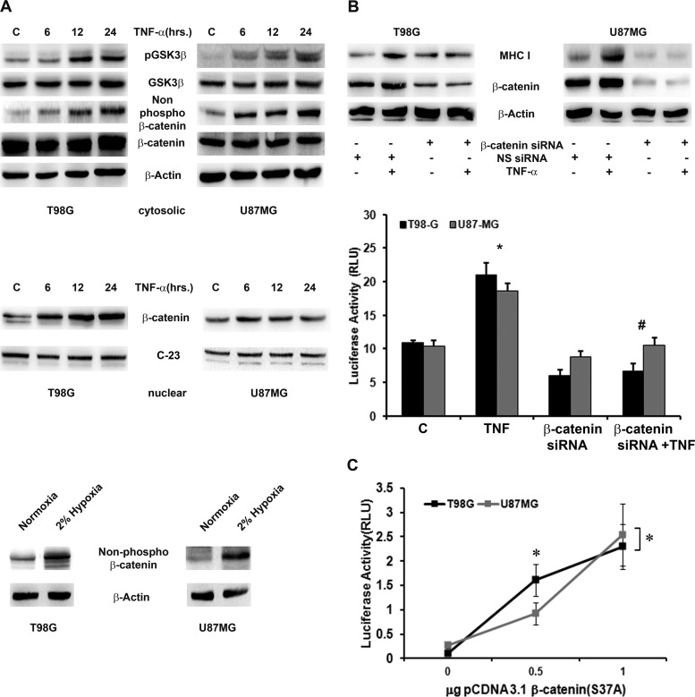
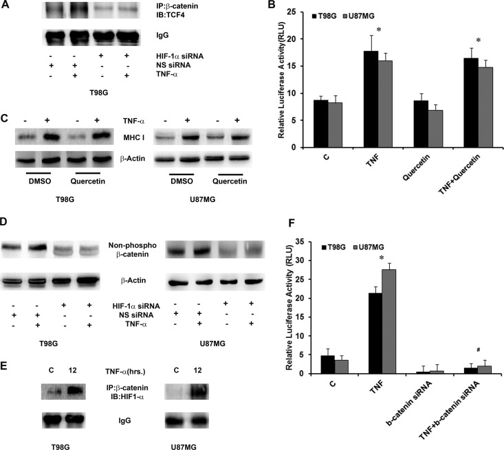
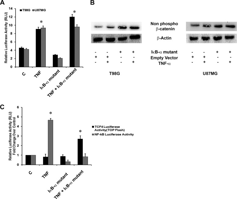
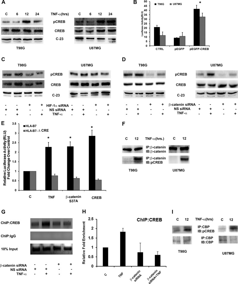
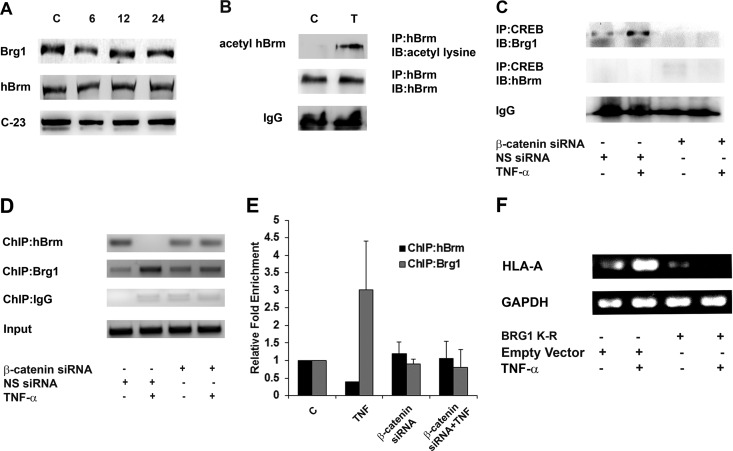
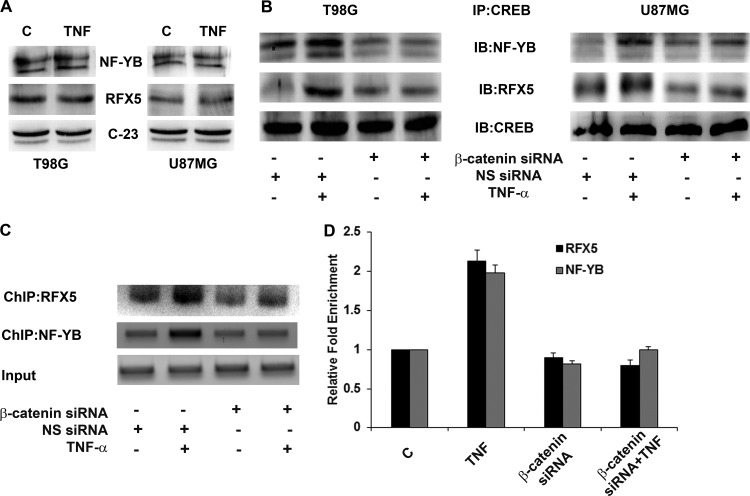
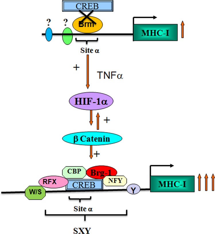
Hypoxia-inducible factor 1α (HIF-1α) plays a crucial role in the progression of glioblastoma multiforme tumors, which are characterized by their effective immune escape mechanisms. As major histocompatibility complex class I (MHC-I) is involved in glioma immune evasion and since HIF-1α is a pivotal link between inflammation and glioma progression, the role of tumor necrosis factor alpha (TNF-α)-induced inflammation in MHC-I gene regulation was investigated. A TNF-α-induced increase in MHC-I expression and transcriptional activation was concurrent with increased HIF-1α, ΝF-κΒ, and β-catenin activities. While knockdown of HIF-1α and β-catenin abrogated TNF-α-induced MHC-I activation, NF-κB had no effect. β-Catenin inhibition abrogated HIF-1α activation and vice versa, and this HIF-1α-β-catenin axis positively regulated CREB phosphorylation. Increased CREB activation was accompanied by its increased association with β-catenin and CBP. Chromatin immunoprecipitation revealed increased CREB enrichment at CRE/site α on the MHC-I promoter in a β-catenin-dependent manner. β-Catenin replaced human Brahma (hBrm) with Brg1 as the binding partner for CREB at the CRE site. The hBrm-to-Brg1 switch is crucial for MHC-I regulation, as ATPase-deficient Brg1 abolished TNF-α-induced MHC-I expression. β-Catenin also increased the association of MHC-I enhanceosome components RFX5 and NF-YB at the SXY module. CREB acts as a platform for assembling coactivators and chromatin remodelers required for MHC-I activation in a HIF-1α/β-catenin-dependent manner.


Figures

Fig 1
Fig 1
TNF-α increases MHC class I expression and transcriptional activation in a HIF-1α-dependent manner. (A) Western blot analysis depicting MHC-I and β2m expression in TNF-α-treated glioma cells. Increased MHC-I heavy chain and β2m expression was also evident by Western blotting. The data are representative of three independent experiments. Blots were reprobed for β-actin to establish equivalent loading. C, control. (B) Surface expression of MHC-I in TNF-α-treated glioma cells as determined by FACS analysis. Shown is a histogram representative of three independent experiments showing the same trend. CTRL, control. (C) TNF-α increases MHC-I transcriptional activity in glioma cells. Luciferase reporter assay of MHC-I transcriptional activity in cells transfected with HLA-B7 reporter constructs and treated with TNF-α for 12 h. RLU, relative light units. (D) TNF-α-induced HIF-1α regulates MHC-I expression. Shown is a Western blot analysis depicting MHC-I levels in glioma cells transfected with HIF-1α siRNA in the presence or absence of TNF-α. The data are representative of three independent experiments. Blots were reprobed for β-actin to establish equivalent loading. NS, nonspecific. (F) TNF-α-induced MHC-I transcriptional activation is HIF-1α dependent. Glioma cells cotransfected either with WT HIF-1α or HIF-1α siRNA and HLA-B7 reporter constructs were treated with TNF-α for 12 h, and a luciferase reporter assay was performed to determine MHC-I promoter activity. Overexpression of WT HIF increases while that of HIF-1α siRNA decreases TNF-α-induced MHC-I promoter activity. (G) Western blot analysis and luciferase reporter assay demonstrating MHC-I expression and activity in cells exposed to 2%hypoxia for 24 h. In panels C, F, and G, the graphs represent normalized firefly luciferase activity. Values represent the means ± the standard errors of the means from three independent experiments. Symbols: *, significant increase over the untreated control; #, significant decrease from TNF-α-treated cells (P < 0.05).
Fig 2
Fig 2
MHC-I expression and transcriptional activation are dependent on β-catenin activation. (A) Increased GSK-3β/β-catenin signaling and nuclear β-catenin expression in TNF-α-treated glioma cells. Shown are Western blot analyses indicating nonphosphorylated and total β-catenin levels in TNF-α-treated cells. The data are representative of three independent experiments. β-Actin and C-23 (nucleolin) levels are shown as loading controls. Western blot analyses also demonstrate nonphosphorylated β-catenin levels in glioma cells treated with 2%hypoxia for 24 h. C, control. (B) TNF-α-induced β-catenin regulates MHC-I expression and activity. Western blot analysis and a luciferase reporter assay were performed with glioma cells either transfected with β-catenin siRNA alone or cotransfected with an HLA-B7 reporter construct in the presence or absence of TNF-α. Blots were reprobed for β-actin to establish equivalent loading. (C) MHC-I transcriptional activity is β-catenin dependent. Glioma cells were cotransfected with an HLA-B7 reporter construct and the indicated amounts of a constitutively active (S37A) β-catenin expression construct. The graph represents relative MHC-I luciferase activity normalized to Renilla luciferase activity. RLU, relative light units. The data shown are representative of three independent experiments. Symbols: *, significant increase over the untreated control; #, significant decrease from TNF-α-treated cells (P < 0.05).
Fig 3
Fig 3
β-Catenin–HIF-1α axis regulates MHC-I expression and transcriptional activation. (A) The TNF-α-induced β-catenin–TCF4 interaction is regulated by HIF-1α. β-Catenin was immunoprecipitated from nuclear lysates as indicated and probed for TCF4. IgG bands were used to normalize levels. IB, immunoblot; NS, nonspecific. (B) MHC I promoter activity and expression are independent of the β-catenin–TCF4 interaction. Following transfection with an HLA-B7 reporter construct, cells were treated with TNF-α in the presence or absence of 10 μM quercetin and a reporter assay was performed. The graph represents relative luciferase activities normalized to control Renilla reporter activity. RLU, relative light units. (C) Western blot analysis was performed to determine MHC I levels in glioma cells treated with different combinations of quercetin and TNF-α. DMSO, dimethyl sulfoxide. (D) Western blot analyses were performed to determine the levels of nonphosphorylated β-catenin in cells transfected with either HIF-1α siRNA or nontargeting siRNA in the presence or absence of TNF-α. The data are representative of three independent experiments. Blots were reprobed for β-actin to establish equivalent loading. (E) TNF-α increases β-catenin–HIF-1α interaction in glioma cells. Nuclear lysates from TNF-α-treated cells were immunoprecipitated with β-catenin and probed with antibody against HIF-1α. IgG levels are shown to establish equivalent loading. A representative blot from two independent experiments with identical results is shown. C, control. (F) Inhibition of β-catenin prevents TNF-α-mediated HIF-1α activation. Following cotransfection with HIF-1α reporter construct and β-catenin siRNA, cells were treated with TNF-α and reporter assay was performed to determine HIF-1α activity. The graph represents relative luciferase activity normalized to Renilla control reporter activity. Data bars represent the means ± the standard errors of the means from three independent experiments. Symbols: *, significant increase over the untreated control, #, significant decrease from TNF-α-treated cells (P < 0.05).
Fig 4
Fig 4
Noninvolvement of NF-κB in TNF-α-induced MHC class I expression and transcriptional activation. (A) TNF-α regulates MHC-I activation in an NF-κB-independent manner. Luciferase reporter assay of MHC-I transcriptional activity in cells cotransfected with HLA-B7 luciferase reporter and IκB-α mutant constructs and treated with TNF-α. C, control; RLU, relative light units. (B) TNF-α-induced NF-κB regulates β-catenin expression. Western blot analyses indicate nonphosphorylated β-catenin levels in TNF-α-treated cells transfected with the IκB-α mutant construct. β-Actin levels are shown as a loading control. (C) TNF-α-induced TCF4 transcriptional activation is NF-κB dependent. Glioma cells cotransfected with the TOP Flash TCF4 reporter construct and the IκB-α mutant construct were treated with TNF-α for 12 h, and a luciferase reporter assay was performed to determine TCF4 promoter activity. The graphs in panels A and C represent normalized luciferase activity over Renilla luciferase values. Values represent the means ± the standard errors of the means from three independent experiments. An asterisk indicates a significant increase over the untreated control.
Fig 5
Fig 5
Involvement of CREB in β-catenin-mediated transactivation of the MHC-I promoter. (A) TNF-α increases CREB phosphorylation in glioma cells. Shown is a Western blot analysis demonstrating CREB levels in cells treated with TNF-α for different intervals of time. C, control. (B) MHC-I transcriptional activation is CREB dependent. Luciferase reporter assay indicating MHC-I activity in glioma cells cotransfected with an HLA-B7 reporter and a pEGFP CREB-WT or an empty pEGFP vector construct. CTRL, control. TNF-α increases CREB phosphorylation in a HIF-1α (C)- and β-catenin (D)-dependent manner. Shown is a Western blot analysis demonstrating CREB phosphorylation in cells transfected with either HIF-1α or β-catenin and nonspecific siRNA and treated with TNF-α. (E) MHC I promoter activity is CRE site dependent. Graphs represent relative luciferase activities normalized to Renilla control reporter activity. Data bars represent the means ± the standard errors of the means of three independent experiments. An asterisk indicates a significant increase over the untreated control (P < 0.05). (F) TNF-α increases β-catenin–CREB interaction in glioma cells. Nuclear extracts from TNF-α-treated cells were immunoprecipitated with β-catenin antibody and then immunoblotted (IB) with CREB antibody. (G and H) ChIP and ChIP-qPCR assays indicating enhanced CREB interaction with CRE in the SXY module of the MHC-I promoter in TNF-α-treated glioma cells. DNA isolated from control and TNF-α-treated cells before and after IP with anti-CREB antibody was amplified with specific primer sets. A representative result from pre-IP (Input) and anti-CREB antibody-immunoprecipitated samples after PCR amplification is shown. ChIP-qPCR data bars represent change in fold enrichment ± SD from 2 independent experiments. NS, nonspecific. (I) IP demonstrating increased interaction between CREB and CBP upon TNF-α treatment. Nuclear extracts from TNF-α-treated cells were immunoprecipitated with CBP antibody and then immunoblotted with pCREB antibody. Panels A, C, and D are representative of three independent experiments. Blots were reprobed for C-23 to establish equivalent loading. Panels F and I are representative blots from two independent experiments with identical results.
Fig 6
Fig 6
Increased association of CREB with Brg1 but not hBrm. (A) TNF-α has no effect on the levels of hBrm and Brg1. Shown is a Western blot analysis indicating nuclear hBrm and Brg1 levels in TNF-α-treated cells. C-23 levels are shown as a loading control (lane C). (B) TNF-α induces acetylation of hBrm. Nuclear extracts from TNF-α-treated cells were immunoprecipitated with anti-hBrm antibody and analyzed for the levels of acetylated hBrm with a pan-acetylated-lysine antibody. IB, immunoblot. (C) TNF-α increases the association between CREB and Brg1 but not hBrm in a β-catenin-dependent manner. Nuclear extracts from cells transfected with β-catenin siRNA and treated with TNF-α were immunoprecipitated with CREB antibody, and an immunoblot analysis was done with Brg1 and hBrm antibodies. Band density was normalized against IgG levels under the same conditions. NS, nonspecific. (D, E) ChIP and ChIP-qPCR analyses indicating the relative changes in hBrm and Brg1 binding levels at the SXY module of the MHC-I promoter upon TNF-α treatment in a β-catenin-dependent manner. A representative gel image of the precleared fraction (Input) and the anti-Brg1- or anti-hBrm antibody-immunoprecipitated sample after PCR amplification is shown. DNA samples immunoprecipitated with the indicated antibodies were also subjected to qPCR along with the diluted input (1%), and the n-fold enrichment was calculated relative to control levels after correction for background signals. qPCR data bars indicate relative changes (n-fold) in enrichment over the control levels ± the standard deviations from two independent sets. (F) Brg1 activity is crucial for MHC-I expression. RT-PCR for MHC-I expression in TNF-α-treated cells transfected with ATPase-deficient Brg1 with a mutation in the ATPase subunit (K-R). GAPDH levels were used as internal controls. All experiments were performed with glioma cell line T98G.
Fig 7
Fig 7
β-Catenin regulates MHC-I enhanceosome formation. (A) TNF-α has no effect on the expression of RFX5 and NF-YB in glioma cells. Shown is a Western blot analysis indicating nuclear RFX5 and NF-YB levels in cells treated with TNF-α for 12 h. C-23 levels are shown as a loading control. (B) TNF-α increases the association of CREB with RFX5 and NF-YB, and this increased association of enhanceosome components is β-catenin dependent. Nuclear extracts from cells transfected with either β-catenin or nontargeting siRNA and treated with TNF-α for 12 h were immunoprecipitated with CREB antibody, and an immunoblot analysis was performed with RFX5 and NF-YB antibodies. Immunoprecipitated CREB levels were used to normalize bands. (C and D) TNF-α regulates the promoter occupancy of RFX5 and NF-YB at the SXY module in a β-catenin-dependent manner in T98G cells. DNA isolated from control and TNF-α-treated cells before and after IP with antibodies as indicated was amplified with specific primer sets. Representative results obtained with pre-IP (Input) and immunoprecipitated samples after PCR amplification are shown. DNA samples immunoprecipitated with the indicated antibodies were also subjected to a qPCR assay along with diluted input (1%), and the relative enrichment was calculated with respect to control levels after correction for background signals. qPCR data bars indicate relative changes in enrichment over control levels ± the standard deviations from two independent sets. The DNA gels and Western blot analyses shown are representative of three independent experiments with similar results. NS, nonspecific.
Fig 8
Fig 8
Model depicting the roles of β-catenin and HIF-1α in TNF-α-mediated MHC-I gene activation. Shown is a schematic representation of the hBrm-to-Brg1 switch at CRE/site α of the MHC-I promoter by HIF-1α. In the absence of inflammation, hBrm occupies the CRE site and prevents the access of CREB to its cognate element. Phosphorylated CREB induced by the TNF-α-triggered HIF-1α–β-catenin axis binds to the CRE site, which is followed by recruitment of CBP and Brg1. TNF-α promotes the formation of the MHC-I enhanceosome by recruiting RFX5 and NF-YB at the CREB/CBP/Brg1 complex. Brg1 mediates an open conformation around the CRE promoter and enhances the HIF-1α-mediated increased induction of MHC-I (arrows). Sites other than CRE/site α drive constitutive MHC-I expression in glioma cells.

Similar articles

Cited by

References

    1. Semenza GL. 2010. Defining the role of hypoxia-inducible factor 1 in cancer biology and therapeutics. Oncogene 29:625–634 - PMC - PubMed
    1. Semenza GL. 2003. Targeting HIF-1 for cancer therapy. Nat. Rev. Cancer 3:721–732 - PubMed
    1. Wang GL, Semenza GL. 1995. Purification and characterization of hypoxia-inducible factor 1. J. Biol. Chem. 270:1230–1237 - PubMed
    1. Wang GL, Jiang BH, Rue EA, Semenza GL. 1995. Hypoxia-inducible factor 1 is a basic-helix-loop-helix-PAS heterodimer regulated by cellular O2 tension. Proc. Natl. Acad. Sci. U. S. A. 92:5510–5514 - PMC - PubMed
    1. Jung Y, Isaacs JS, Lee S, Trepel J, Liu ZG, Neckers L. 2003. Hypoxia-inducible factor induction by tumour necrosis factor in normoxic cells requires receptor-interacting protein-dependent nuclear factor kappa B activation. Biochem. J. 370:1011–1017 - PMC - PubMed

Publication types

MeSH terms

Substances