

Neurological soft signs are not "soft" in brain structure and functional networks: evidence from ALE meta-analysis
- PMID: 23671197
- PMCID: PMC3984512
- DOI: 10.1093/schbul/sbt063
Neurological soft signs are not "soft" in brain structure and functional networks: evidence from ALE meta-analysis
Abstract
Background: Neurological soft signs (NSS) are associated with schizophrenia and related psychotic disorders. NSS have been conventionally considered as clinical neurological signs without localized brain regions. However, recent brain imaging studies suggest that NSS are partly localizable and may be associated with deficits in specific brain areas.
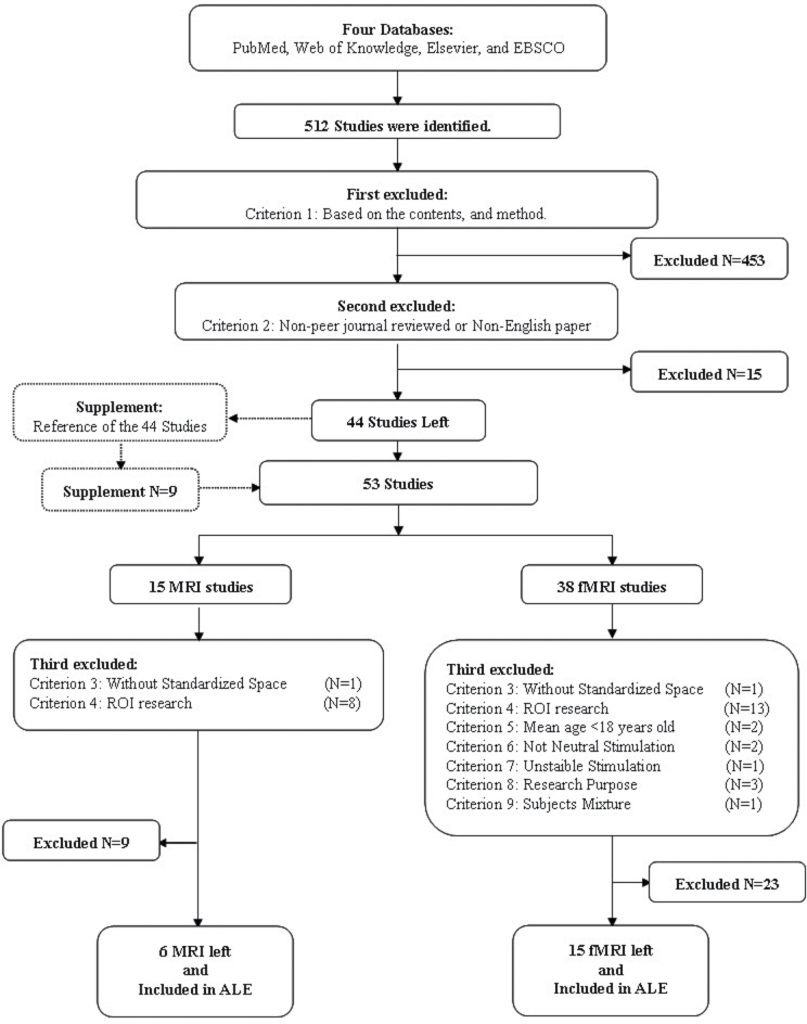
Method: We conducted an activation likelihood estimation meta-analysis to quantitatively review structural and functional imaging studies that evaluated the brain correlates of NSS in patients with schizophrenia and other psychotic disorders. Six structural magnetic resonance imaging (sMRI) and 15 functional magnetic resonance imaging (fMRI) studies were included.
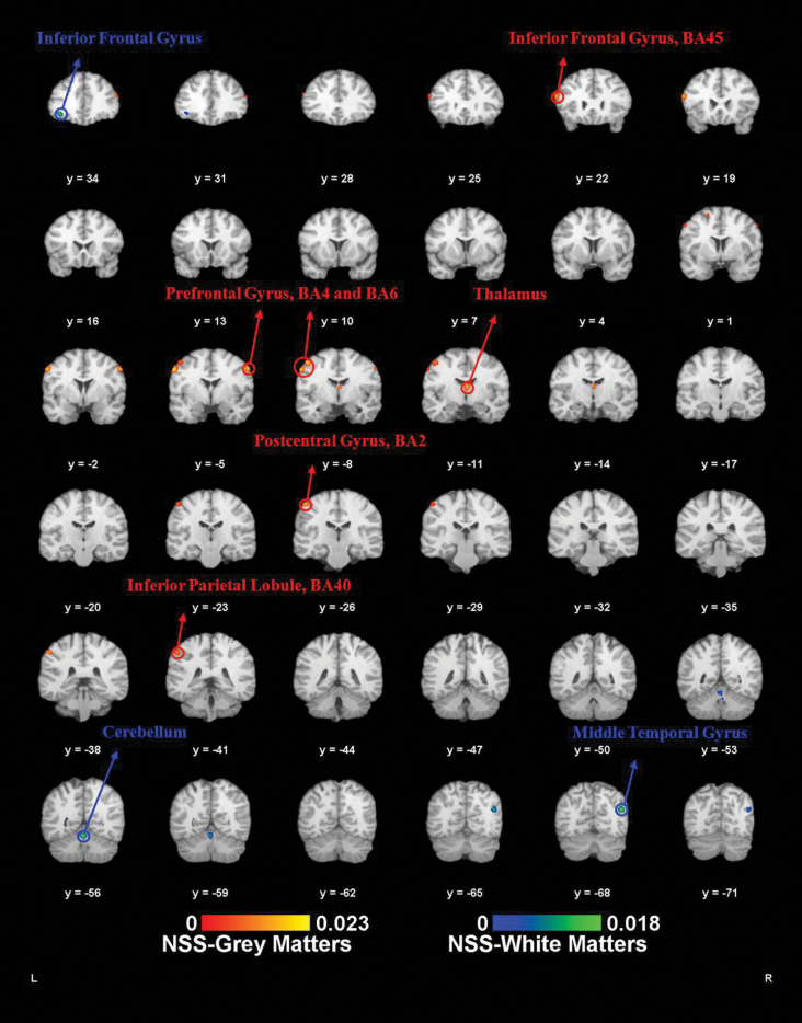
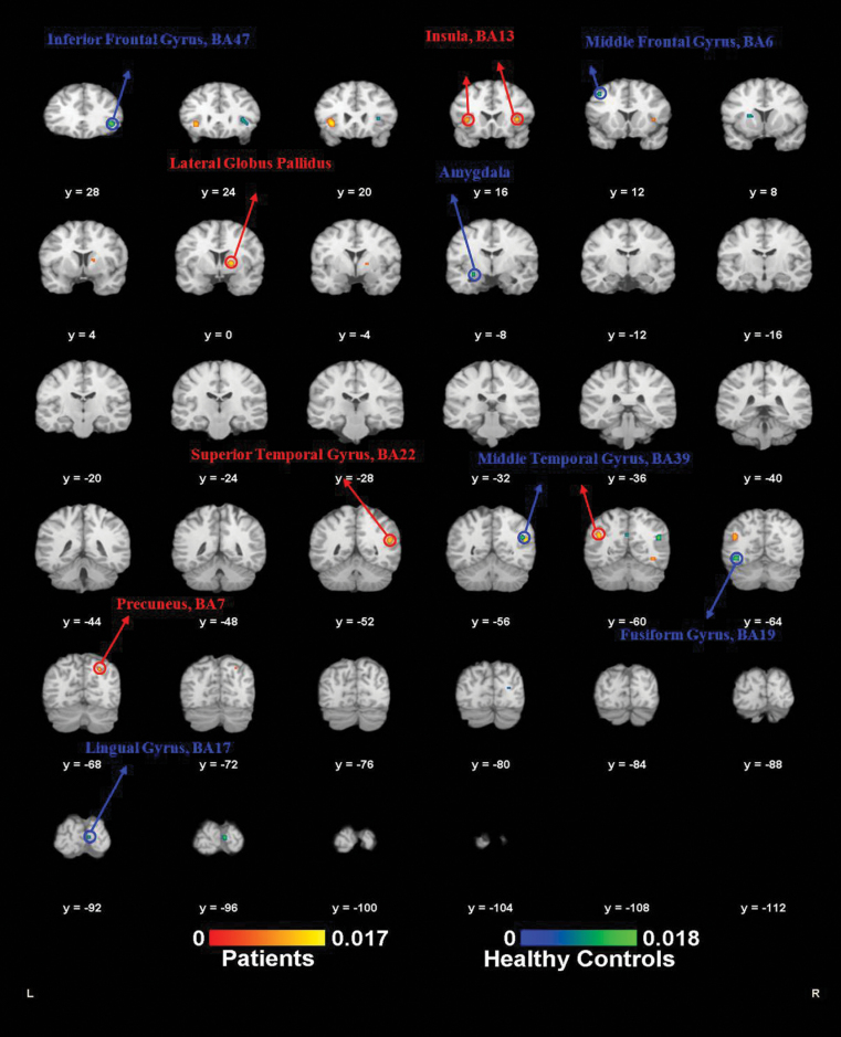
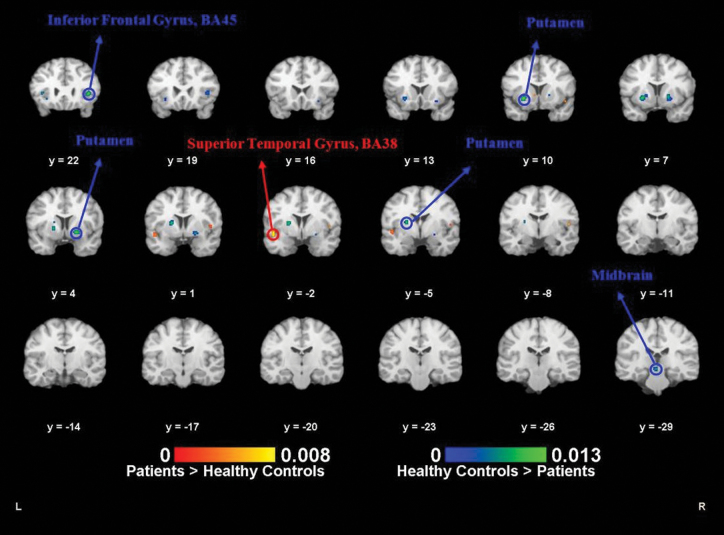
Results: The results from meta-analysis of the sMRI studies indicated that NSS were associated with atrophy of the precentral gyrus, the cerebellum, the inferior frontal gyrus, and the thalamus. The results from meta-analysis of the fMRI studies demonstrated that the NSS-related task was significantly associated with altered brain activation in the inferior frontal gyrus, bilateral putamen, the cerebellum, and the superior temporal gyrus.
Conclusions: Our findings from both sMRI and fMRI meta-analyses further support the conceptualization of NSS as a manifestation of the "cerebello-thalamo-prefrontal" brain network model of schizophrenia and related psychotic disorders.
Keywords: activation; brain; estimation; imaging; likelihood; meta-analysis; neurological; psychosis; schizophrenia; signs; soft.
Figures
References
-
- Tsuang MT, Gilbertson MW, Faraone SV. The genetics of schizophrenia. Current knowledge and future directions. Schizophr Res. 1991;4:157–171 - PubMed
-
- Tsuang MT, Faraone SV, Lyons MJ. Identification of the phenotype in psychiatric genetics. Eur Arch Psychiatry Clin Neurosci. 1993;243:131–142 - PubMed
-
- Chan RC, Gottesman II. Neurological soft signs as candidate endophenotypes for schizophrenia: a shooting star or a Northern star? Neurosci Biobehav Rev. 2008;32:957–971 - PubMed
-
- Smith RC, Hussain MI, Chowdhury SA, Stearns A. Stability of neurological soft signs in chronically hospitalized schizophrenic patients. J Neuropsychiatry Clin Neurosci. 1999;11:91–96 - PubMed
Publication types
MeSH terms
LinkOut - more resources
Full Text Sources
Other Literature Sources
Medical
