. 2013 Jul;154(7):2296-307.
doi: 10.1210/en.2012-2077. Epub 2013 May 13.

5α-reductase inhibition suppresses testosterone-induced initial regrowth of regressed xenograft prostate tumors in animal models

Affiliations

5α-reductase inhibition suppresses testosterone-induced initial regrowth of regressed xenograft prostate tumors in animal models

Khalid Z Masoodi et al. Endocrinology. 2013 Jul.

Abstract

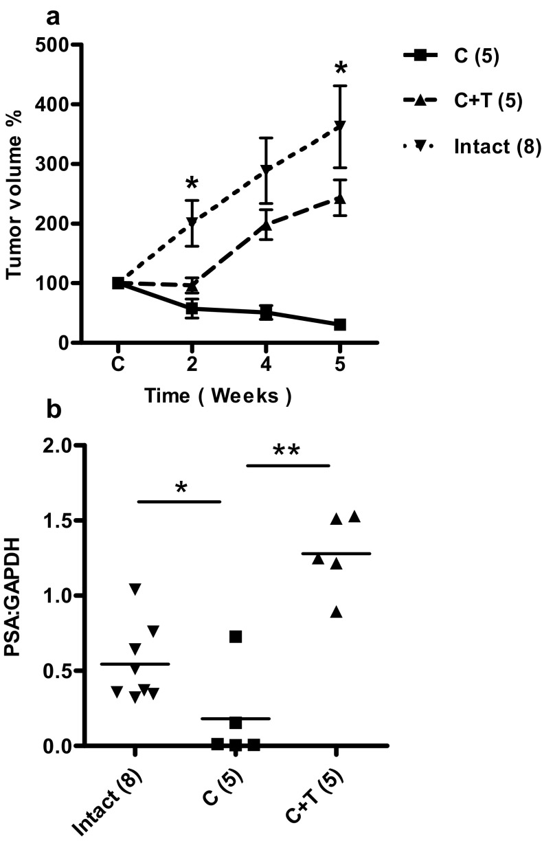
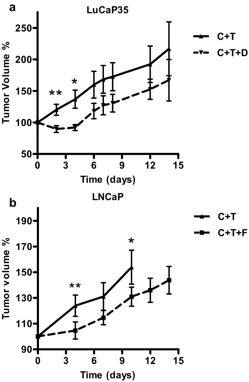
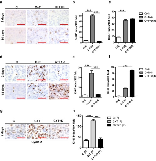
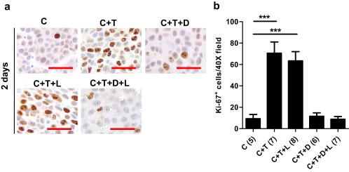
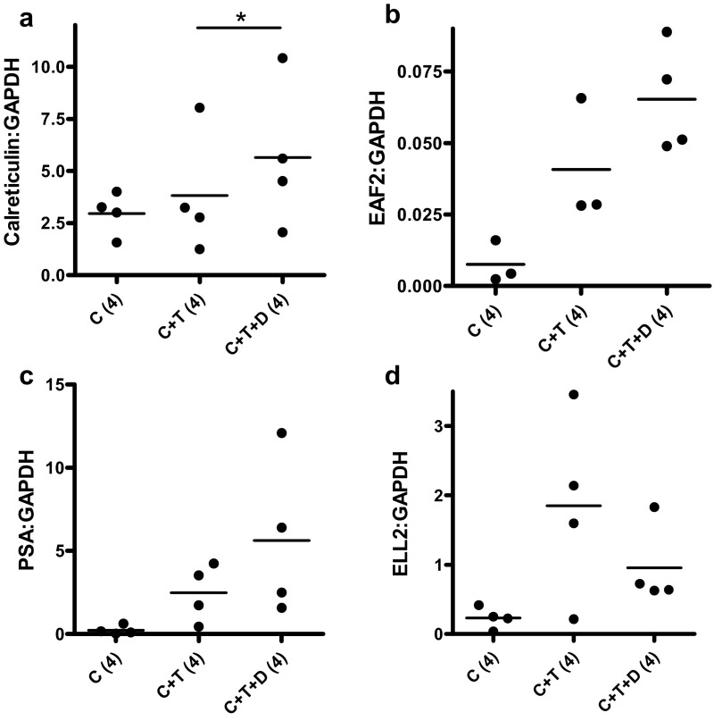
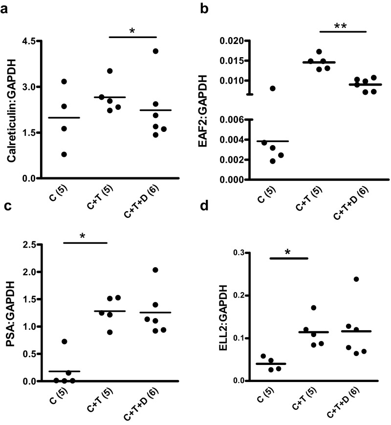
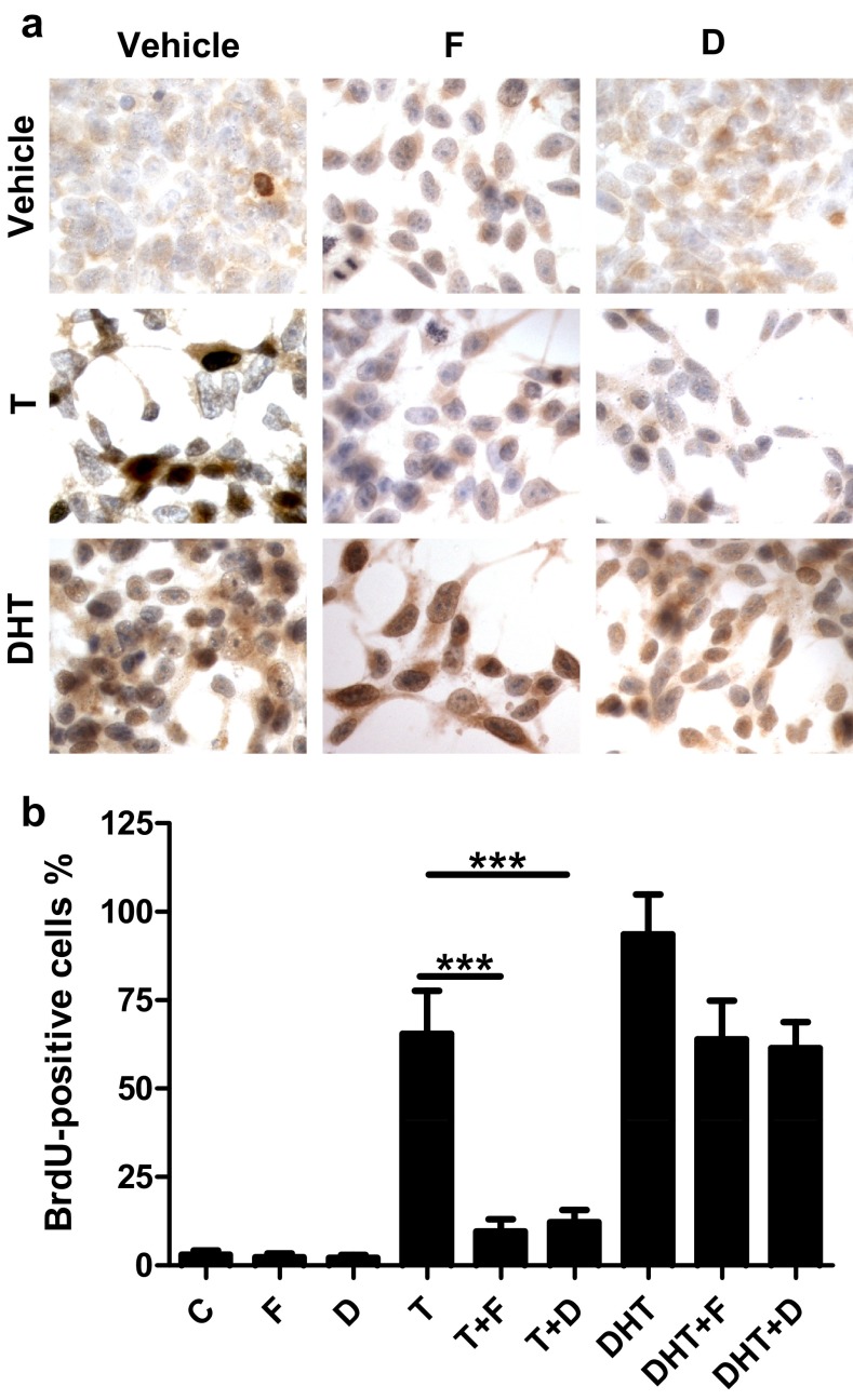
Androgen deprivation therapy (ADT) is the standard treatment for patients with prostate-specific antigen progression after treatment for localized prostate cancer. An alternative to continuous ADT is intermittent ADT (IADT), which allows recovery of testosterone during off-cycles to stimulate regrowth and differentiation of the regressed prostate tumor. IADT offers patients a reduction in side effects associated with ADT, improved quality of life, and reduced cost with no difference in overall survival. Our previous studies showed that IADT coupled with 5α-reductase inhibitor (5ARI), which blocks testosterone conversion to DHT could prolong survival of animals bearing androgen-sensitive prostate tumors when off-cycle duration was fixed. To further investigate this clinically relevant observation, we measured the time course of testosterone-induced regrowth of regressed LuCaP35 and LNCaP xenograft tumors in the presence or absence of a 5ARI. 5α-Reductase inhibitors suppressed the initial regrowth of regressed prostate tumors. However, tumors resumed growth and were no longer responsive to 5α-reductase inhibition several days after testosterone replacement. This finding was substantiated by bromodeoxyuridine and Ki67 staining of LuCaP35 tumors, which showed inhibition of prostate tumor cell proliferation by 5ARI on day 2, but not day 14, after testosterone replacement. 5α-Reductase inhibitors also suppressed testosterone-stimulated proliferation of LNCaP cells precultured in androgen-free media, suggesting that blocking testosterone conversion to DHT can inhibit prostate tumor cell proliferation via an intracrine mechanism. These results suggest that short off-cycle coupled with 5α-reductase inhibition could maximize suppression of prostate tumor growth and, thus, improve potential survival benefit achieved in combination with IADT.


Figures

Figure 1.
Figure 1.
Experimental protocols. Tumor-bearing SCID/Nude mice were castrated and subjected to 3 different protocols, a, b, and c. Animals in protocol A were followed for 14 days before randomization into 3 subgroups: implantation of testosterone (C+T), testosterone and dutasteride (C+T+D), and no intervention (C). Tumor volume was measured daily for the initial 7 days of treatment and on alternate days thereafter. Tumors were collected at days 2, 4, and 14 for IHC and qPCR (n = 4 for each group). In protocol B, mice were followed for 10 days before randomization and implantation (n = 7 for each group). Testosterone and dutasteride pellets were removed 4 days after implantation. Ten days after pellet removal, testosterone and/or dutasteride pellets were again implanted. Tumors were then collected for IHC 2 days after the pellet implantation. In protocol C, mice were followed for 10 days after castration and randomized into 5 groups, consisting of C (n = 5), C+T (n = 7), C+T+D (n = 6), C+T+L (n = 8), and C+T+D+L (n = 7). Tumors were collected at day 2 for IHC.
Figure 2.
Figure 2.
Response of LuCaP35 xenograft tumors to androgen manipulation. Panel A, Effect of castration on tumor volume in castrated (C), castrated plus testosterone replacement (C+T) and testes-intact control animals. Values expressed as percentage of original volume (200 mm3). Panel B, Effect of androgen replacement on PSA mRNA expression level relative to GAPDH. Values are presented as mean ± SEM. *, P < .05; **, P < .01. Number of animals in each group is shown in parentheses.
Figure 3.
Figure 3.
Response of LuCaP35 and LNCaP xenograft tumors to 5ARIs- dutasteride and finasteride. Panel A, Effect of dutasteride on LuCaP35 tumor volume. Tumor volume in castrated animals with testosterone replacement (C + T) (n = 5), and animals treated with testosterone replacement along with dutasteride (C + T + D) (n = 5) at indicated time points. Panel B, Effect of finasteride on LNCaP tumor volume. Tumor volume in castrated animals with testosterone replacement (C + T) (n = 6) and in castrated animals with testosterone replacement along with finasteride (C + T + F) (n = 8) at indicated time points. Tumor volume was determined as the percentage of tumor volume at time of implantation of T, D, and/or F pellets. *, P < .05; **, P < .01.
Figure 4.
Figure 4.
Effects of dutasteride on cellular proliferation in LuCaP35 and LNCaP tumors. Panel A, BrdU labeling in transverse sections of LuCaP35 xenograft tumors from C, C+T, and C+T+D mice 2 and 14 days after testosterone implantation. Panels B and C, Quantification of BrdU label-retaining cells in LuCaP35 tumors at days 2 and 14 after castration. Panel D, Ki-67 immunostaining in transverse sections of LuCaP35 xenograft tumors from C, C+T, and C+T+D mice 2 and 14 days after testosterone implantation. Panels E and F, Quantification of Ki-67–positive cells in LuCaP35 tumors at days 2 and 14 after castration. Data represent the average of 4 mice per group, which is indicated in parentheses. Panel G, Ki-67 immunostaining in transverse sections of LNCaP xenograft tumors from C, C+T, and C+T+D mice collected at day 2 of the second cycle of testosterone/dutasteride implantation. Panel H, Quantification of Ki-67–positive cells in LNCaP tumors at day 2 of the second cycle after testosterone replacement. Number of animals in each group is shown in parentheses. Scale bars, 50 μm. ***, P < .0001.
Figure 5.
Figure 5.
Effect of aromatase inhibitor letrozole on cellular proliferation marker Ki-67 expression in LNCaP tumor xenografts. A, Ki-67 immunostaining in transverse sections of xenograft tumors from C, C+T, C+T+D, C+T+L, and C+T+D+L mice 2 days after testosterone replacement. Panel B, Quantification of Ki-67–positive cells in LNCaP tumors at day 2 post testosterone replacement. Error bars represent SEM. Number of animals in each group is shown in parentheses. ***, P < .0001.
Figure 6.
Figure 6.
Effect of dutasteride on the expression of various androgen-responsive genes in LuCaP35 tumors 4 days after testosterone implantation. Data shown are qPCR analysis of androgen-responsive genes, calreticulin, EAF2, PSA, and ELL2, in C, C+T, and C+T+D LuCaP35 xenograft tumors. Data were normalized to GAPDH. Number of animals in each group is shown in parentheses. Each dot represents a single sample; the line depicts the mean. *, P < .05.
Figure 7.
Figure 7.
Effect of dutasteride on the expression of various androgen-responsive genes in LuCaP35 tumors 14 days after testosterone implantation. Data shown are qPCR analysis of androgen-responsive genes, calreticulin, EAF2, PSA, and ELL2, in C, C+T, and C+T+D LuCaP35 xenograft tumors. Data were normalized to GAPDH. Number of animals in each group is shown in parentheses. Each dot represents a single sample; the line depicts the mean. *, P < .05.
Figure 8.
Figure 8.
Effects of dutasteride or finasteride on testosterone-stimulated cellular proliferation in cultured LNCaP cells. Panel A, Representative images of BrdU immunostaining in LNCaP cells precultured in charcoal-stripped media for 48 hours and subsequently cultured for 3 days under the following conditions: dimethylsulfoxide vehicle control (C), 5nM finasteride (F), 5nM dutasteride (D), 1nM testosterone (T), 1nM testosterone plus 5nM finasteride (T+F), 1nM testosterone plus 5nM dutasteride (T+D), 1nM DHT, 1nM DHT plus 5nM finasteride (DHT+F), 1nM DHT plus 5nM dutasteride (DHT+D). Panel B, Quantification of BrdU-positive cells. Data are representative of 5 independent experiments. Error bars represent SEM. ***, P < .0001.

References

    1. Coffey DS, Shimazaki J, Williams-Ashman HG. Polymerization of deoxyribonucleotides in relation to androgen-induced prostatic growth. Arch Biochem Biophys. 1968;124:184–198 - PubMed
    1. Huggins Endocrine-induced regression of cancers. Cancer Research. 1967;27:1925–1930 - PubMed
    1. Yang Q, Fung KM, Day WV, Kropp BP, Lin HK. Androgen receptor signaling is required for androgen-sensitive human prostate cancer cell proliferation and survival. Cancer Cell Int. 2005;5:8. - PMC - PubMed
    1. Thomas LN, Douglas RC, Lazier CB, et al. Levels of 5α-reductase type 1 and type 2 are increased in localized high grade compared to low grade prostate cancer. J Urol. 2008;179:147–151 - PubMed
    1. Zhou ZX, Lane MV, Kemppainen JA, French FS, Wilson EM. Specificity of ligand-dependent androgen receptor stabilization: receptor domain interactions influence ligand dissociation and receptor stability. Mol Endocrinol. 1995;9:208–218 - PubMed

Publication types

MeSH terms