. 2013 Jun 28;288(26):19269-79.
doi: 10.1074/jbc.M112.445445. Epub 2013 May 13.

Impaired very long-chain acyl-CoA β-oxidation in human X-linked adrenoleukodystrophy fibroblasts is a direct consequence of ABCD1 transporter dysfunction

Affiliations

Impaired very long-chain acyl-CoA β-oxidation in human X-linked adrenoleukodystrophy fibroblasts is a direct consequence of ABCD1 transporter dysfunction

Christoph Wiesinger et al. J Biol Chem. .

Abstract

X-linked adrenoleukodystrophy (X-ALD), an inherited peroxisomal disorder, is caused by mutations in the ABCD1 gene encoding the peroxisomal ATP-binding cassette (ABC) transporter ABCD1 (adrenoleukodystrophy protein, ALDP). Biochemically, X-ALD is characterized by an accumulation of very long-chain fatty acids and partially impaired peroxisomal β-oxidation. In this study, we used primary human fibroblasts from X-ALD and Zellweger syndrome patients to investigate the peroxisomal β-oxidation defect. Our results show that the degradation of C26:0-CoA esters is as severely impaired as degradation of unesterified very long-chain fatty acids in X-ALD and is abolished in Zellweger syndrome. Interestingly, the β-oxidation rates for both C26:0-CoA and C22:0-CoA were similarly affected, although C22:0 does not accumulate in patient fibroblasts. Furthermore, we show that the β-oxidation defect in X-ALD is directly caused by ABCD1 dysfunction as blocking ABCD1 function with a specific antibody reduced β-oxidation to levels observed in X-ALD fibroblasts. By quantification of mRNA and protein levels of the peroxisomal ABC transporters and by blocking with specific antibodies, we found that residual β-oxidation activity toward C26:0-CoA in X-ALD fibroblasts is mediated by ABCD3, although the efficacy of ABCD3 appeared to be much lower than that of ABCD1. Finally, using isolated peroxisomes, we show that β-oxidation of C26:0-CoA is independent of additional CoA but requires a cytosolic factor of >10-kDa molecular mass that is resistant to N-ethylmaleimide and heat inactivation. In conclusion, our findings in human cells suggest that, in contrast to yeast cells, very long-chain acyl-CoA esters are transported into peroxisomes by ABCD1 independently of additional synthetase activity.

Keywords: ABC Transporter; ABCD1; ABCD3; Adrenoleukodystrophy Protein; Fatty Acid Transport; Metabolic Diseases; Peroxisomes; VLCFA; X-ALD; β-Oxidation.


Figures
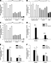
FIGURE 1.
FIGURE 1.
β-Oxidation of free VLCFAs and VLCFA-CoA esters in homogenates from human fibroblasts. Cultured fibroblasts from healthy controls, X-ALD patients, or ZWS patients were homogenized in isoosmotic buffer, and β-oxidation activities of C26:0 (A), C16:0 (B), and C26:0-CoA (C) were determined in postmitochondrial supernatants. D, C26:0-CoA β-oxidation activity of homogenates from healthy and X-ALD fibroblasts was measured in the absence (light gray) or presence (black) of exogenous ATP and in the presence of ATP after preincubation with apyrase (dark gray). E, C26:0-CoA β-oxidation activity was measured upon preincubation of homogenates with a specific α-ABCD1 antibody (Ab) (dark gray), an unspecific antibody (light gray), or no addition of antibody (black). F, C26:0 free fatty acid β-oxidation activity was determined upon antibody inhibition as described in E. The rate of β-oxidation is expressed as pmol of labeled acetate released/min/mg of protein of homogenate added. Values given are means ± S.D. (error bars) of three measurements in one cell line (A–C) or means ± S.D. (error bars) of at least three different cell lines (D–F) (*, p < 0.05; ***, p < 0.001; n.s., not significant).
FIGURE 2.
FIGURE 2.
β-Oxidation of C22:0-CoA is defective in X-ALD but does not lead to accumulation of C22:0. A, fibroblasts were grown to confluence, and after extraction and hydrolysis, the levels of C22:0, C24:0, and C26:0 fatty acids were measured by GC-MS and are depicted as -fold increase over healthy control values. Values represent data normalized to the healthy controls and are given as means ± S.D. (error bars) of at least three different cell lines. B, β-oxidation activities of C22:0-CoA in fibroblast homogenates were determined similarly as described in Fig. 1. C, C22:0-CoA β-oxidation was determined upon inhibition of ABCD1 function by preincubation with an α-ABCD1 antibody (Ab) (dark gray), an unspecific antibody (light gray), or no addition of antibody (black). Values represent means ± S.D. (error bars) of three measurements (**, p < 0.01; ***, p < 0.001; n.s., not significant).
FIGURE 3.
FIGURE 3.
The abundance of the three peroxisomal ABC transporters in human fibroblasts and the relative contribution of ABCD3 to C26:0-CoA degradation. A, mRNA was isolated from cultured human fibroblasts of healthy controls, X-ALD patients, and ZWS patients. The amount of mRNA encoding ABCD1, ABCD2, and ABCD3 was quantified by quantitative RT-PCR and is presented as relative copy number normalized to the housekeeping gene HPRT (means ± S.D. (error bars) of three independent RNA preparations). Note that ABCD2 is virtually absent as a different scale was used for ABCD2. B, the relative abundance of ABCD1 and ABCD3 at the protein level was determined by comparing the immunoblot signals of ABCD1 and ABCD3 with a fusion protein containing the epitopes recognized by the respective antibodies. The fusion construct consisted of full-length ABCD1 (as the exact epitope of the antibody is not known), the humanized epitope of the α-ABCD3 antibody (Ab), and an EGFP tag. Equal amounts of fibroblast homogenates were separated by SDS-PAGE and subjected to Western blot analysis, and the signals of either ABCD1 or ABCD3 were compared with serial dilutions of fusion protein applied in parallel lanes. The indicated values below each panel represent the quantification (arbitrary units) relative to the fusion protein (n.q., not quantified). C, the fraction of C26:0-CoA β-oxidation activity mediated by ABCD3 was obtained by measuring C26:0-CoA β-oxidation in homogenates from X-ALD fibroblasts and determining the effects upon addition of ATP (lane 2) or preincubation with either α-ABCD3 antibody (lane 3) or α-ABCD3 antibody blocked by an excess of the immunizing peptide (pept) (lane 4). Values represent means ± S.D. (error bars) of specific activity of at least three independent cell lines. D and E, summary of the ABCD1 and ABCD3 mRNA (D) and protein (E) levels in healthy fibroblasts. Values represent means ± S.D. (error bars) of five healthy controls. F, the relative contribution of ABCD1 and ABCD3 to C26:0-CoA β-oxidation activity was estimated by determination of the inhibitory effect exerted by α-ABCD1 or α-ABCD3 antibodies. The values shown represent the decrease obtained by inhibition with antibodies as determined in Fig. 1E for ABCD1 (in healthy control fibroblasts) or Fig. 3 C for ABCD3 (**, p < 0.01; ***, p < 0.001). AA, amino acids.
FIGURE 4.
FIGURE 4.
A cytosolic factor is needed for functional β-oxidation of VLCFA in purified peroxisomes. A, fibroblast homogenates were subfractionated by OptiPrep density gradient centrifugation. The distribution of the different organelles in the gradient is shown by Western blot analysis using the respective marker enzymes ABCD1 and ABCD3 (peroxisomes), ATPase (mitochondria), GRP78 (endoplasmic reticulum), LAMP2 (lysosomes), and lactate dehydrogenase (LDH) (cytosol). B, C26:0-CoA β-oxidation in the peroxisomal fraction (pool of fractions without detectable levels of other marker proteins) of healthy controls in the presence and absence of a cytosolic fraction (supernatant of 200,000 × g). Separation of the cytosolic fraction into low (<10-kDa) and high (>10-kDa) molecular mass molecules was carried out by ultrafiltration with a cutoff size of 10 kDa. C, peroxisomes and cytosol of either X-ALD or healthy fibroblasts were combined crosswise, and C26:0-CoA β-oxidation rates were determined. D, C26:0 β-oxidation was determined in peroxisomal fractions derived from healthy fibroblasts supplemented with cytosolic fractions inactivated (inact.) either by NEM or heat treatment for 10 min as indicated. Cytosol derived from ZWS fibroblasts was used to exclude the possibility of leakage of peroxisomal enzymes into the cytosol during homogenization. β-Oxidation rates in the presence of cytosol were taken as reference (100%) for each individual cell line. Values represent means ± S.D. (error bars) when comparing at least three different cell lines (**, p < 0.01; ***, p < 0.001; n.s., not significant).
FIGURE 5.
FIGURE 5.
Degradation of C26:0-CoA is independent of an additional synthetase activity. Peroxisomal fractions derived from either healthy or X-ALD fibroblasts were supplemented with NEM-inactivated cytosolic fractions lacking cofactors of less than 10 kDa, and C26:0-CoA β-oxidation activity was determined in the presence (black) or absence (gray) of ATP (A) and upon preincubation with either an α-ABCD1 antibody (Ab) (dark gray), an unspecific antibody (light gray), or no addition of antibody (black) (B). C, a peroxisomal fraction derived from healthy fibroblasts was supplemented with an NEM-inactivated cytosolic fraction lacking cofactors of less than 10 kDa, and β-oxidation activities of free C26:0 fatty acids or the respective C26:0-CoA esters were determined in the presence or absence of CoA. Dependence on NAD+ was used as a control for the permeability of the peroxisomal membrane for cofactors. β-Oxidation rates in the presence of all cofactors were set as 100% for C26:0 and C26:0-CoA, respectively. Values represent means ± S.D. (error bars) of at least three different cell lines (**, p < 0.01; ***, p < 0.001; n.s., not significant).

References

    1. Bezman L., Moser A. B., Raymond G. V., Rinaldo P., Watkins P. A., Smith K. D., Kass N. E., Moser H. W. (2001) Adrenoleukodystrophy: incidence, new mutation rate, and results of extended family screening. Ann. Neurol. 49, 512–517 - PubMed
    1. Ferrer I., Aubourg P., Pujol A. (2010) General aspects and neuropathology of X-linked adrenoleukodystrophy. Brain Pathol. 20, 817–830 - PMC - PubMed
    1. Kemp S., Berger J., Aubourg P. (2012) X-linked adrenoleukodystrophy: clinical, metabolic, genetic and pathophysiological aspects. Biochim. Biophys. Acta 1822, 1465–1474 - PubMed
    1. Moser A. B., Moser H. W. (1999) The prenatal diagnosis of X-linked adrenoleukodystrophy. Prenat. Diagn. 19, 46–48 - PubMed
    1. Schutgens R. B., Bouman I. W., Nijenhuis A. A., Wanders R. J., Frumau M. E. (1993) Profiles of very-long-chain fatty acids in plasma, fibroblasts, and blood cells in Zellweger syndrome, X-linked adrenoleukodystrophy, and rhizomelic chondrodysplasia punctata. Clin. Chem. 39, 1632–1637 - PubMed

Publication types

MeSH terms