

Autophagosomal Syntaxin17-dependent lysosomal degradation maintains neuronal function in Drosophila
- PMID: 23671310
- PMCID: PMC3653357
- DOI: 10.1083/jcb.201211160
Autophagosomal Syntaxin17-dependent lysosomal degradation maintains neuronal function in Drosophila
Abstract
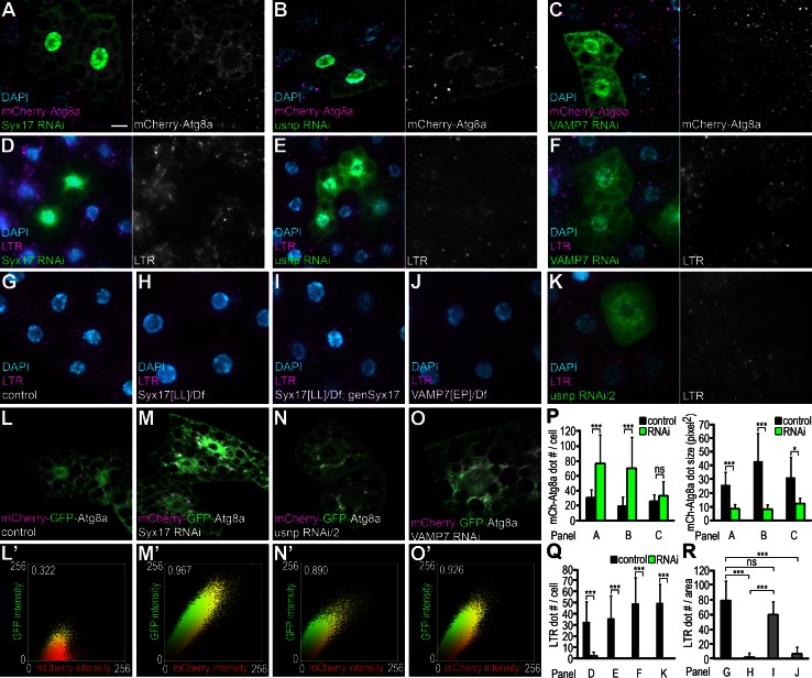
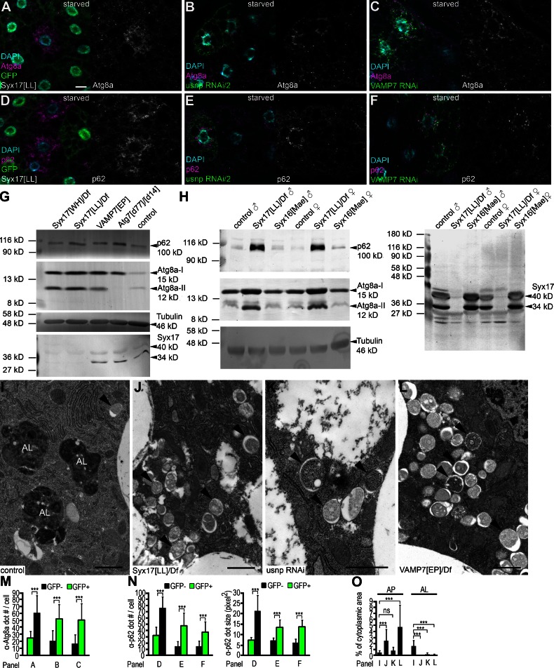
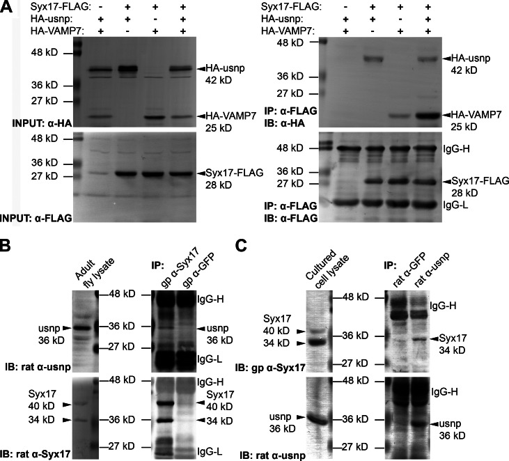
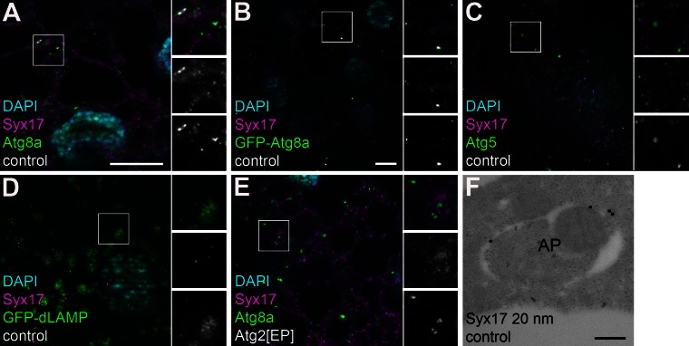
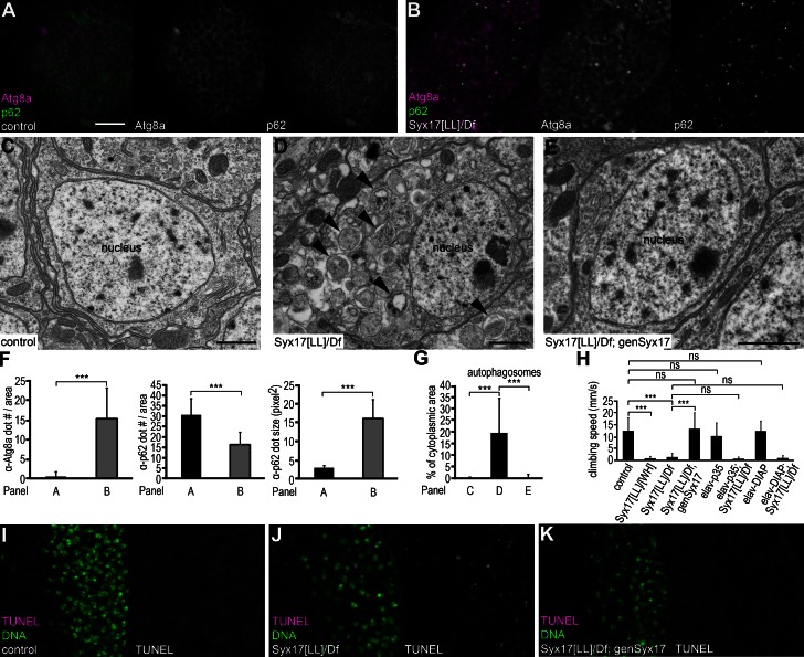
During autophagy, phagophores capture portions of cytoplasm and form double-membrane autophagosomes to deliver cargo for lysosomal degradation. How autophagosomes gain competence to fuse with late endosomes and lysosomes is not known. In this paper, we show that Syntaxin17 is recruited to the outer membrane of autophagosomes to mediate fusion through its interactions with ubisnap (SNAP-29) and VAMP7 in Drosophila melanogaster. Loss of these genes results in accumulation of autophagosomes and a block of autolysosomal degradation during basal, starvation-induced, and developmental autophagy. Viable Syntaxin17 mutant adults show large-scale accumulation of autophagosomes in neurons, severe locomotion defects, and premature death. These mutant phenotypes cannot be rescued by neuron-specific inhibition of caspases, suggesting that caspase activation and cell death do not play a major role in brain dysfunction. Our findings reveal the molecular mechanism underlying autophagosomal fusion events and show that lysosomal degradation and recycling of sequestered autophagosome content is crucial to maintain proper functioning of the nervous system.
Figures
Comment in
-
Route to destruction: autophagosomes SNARE lysosomes.J Cell Biol. 2013 May 13;201(4):495-7. doi: 10.1083/jcb.201304065. J Cell Biol. 2013. PMID: 23671308 Free PMC article.
-
A genetic model with specifically impaired autophagosome-lysosome fusion.Autophagy. 2013 Aug;9(8):1251-2. doi: 10.4161/auto.25470. Epub 2013 Jun 25. Autophagy. 2013. PMID: 23819962 Free PMC article.
Similar articles
-
A genetic model with specifically impaired autophagosome-lysosome fusion.Autophagy. 2013 Aug;9(8):1251-2. doi: 10.4161/auto.25470. Epub 2013 Jun 25. Autophagy. 2013. PMID: 23819962 Free PMC article.
-
Route to destruction: autophagosomes SNARE lysosomes.J Cell Biol. 2013 May 13;201(4):495-7. doi: 10.1083/jcb.201304065. J Cell Biol. 2013. PMID: 23671308 Free PMC article.
-
Evolutionarily conserved role and physiological relevance of a STX17/Syx17 (syntaxin 17)-containing SNARE complex in autophagosome fusion with endosomes and lysosomes.Autophagy. 2013 Oct;9(10):1642-6. doi: 10.4161/auto.25684. Epub 2013 Jul 22. Autophagy. 2013. PMID: 24113031 Review.
-
A voltage-gated calcium channel regulates lysosomal fusion with endosomes and autophagosomes and is required for neuronal homeostasis.PLoS Biol. 2015 Mar 26;13(3):e1002103. doi: 10.1371/journal.pbio.1002103. eCollection 2015 Mar. PLoS Biol. 2015. PMID: 25811491 Free PMC article.
-
The multi-functional SNARE protein Ykt6 in autophagosomal fusion processes.Cell Cycle. 2019 Mar-Apr;18(6-7):639-651. doi: 10.1080/15384101.2019.1580488. Epub 2019 Mar 17. Cell Cycle. 2019. PMID: 30836834 Free PMC article. Review.
Cited by
-
Autophagosome-lysosome fusion in neurons requires INPP5E, a protein associated with Joubert syndrome.EMBO J. 2016 Sep 1;35(17):1853-67. doi: 10.15252/embj.201593148. Epub 2016 Jun 23. EMBO J. 2016. PMID: 27340123 Free PMC article.
-
V-ATPase controls tumor growth and autophagy in a Drosophila model of gliomagenesis.Autophagy. 2021 Dec;17(12):4442-4452. doi: 10.1080/15548627.2021.1918915. Epub 2021 May 12. Autophagy. 2021. PMID: 33978540 Free PMC article.
-
New insights regarding SNARE proteins in autophagosome-lysosome fusion.Autophagy. 2021 Oct;17(10):2680-2688. doi: 10.1080/15548627.2020.1823124. Epub 2020 Sep 24. Autophagy. 2021. PMID: 32924745 Free PMC article. Review.
-
Molecular Mechanism and Regulation of Autophagy and Its Potential Role in Epilepsy.Cells. 2022 Aug 23;11(17):2621. doi: 10.3390/cells11172621. Cells. 2022. PMID: 36078029 Free PMC article. Review.
-
An in vivo RNAi screen uncovers the role of AdoR signaling and adenosine deaminase in controlling intestinal stem cell activity.Proc Natl Acad Sci U S A. 2020 Jan 7;117(1):464-471. doi: 10.1073/pnas.1900103117. Epub 2019 Dec 18. Proc Natl Acad Sci U S A. 2020. PMID: 31852821 Free PMC article.
References
-
- Axe E.L., Walker S.A., Manifava M., Chandra P., Roderick H.L., Habermann A., Griffiths G., Ktistakis N.T. 2008. Autophagosome formation from membrane compartments enriched in phosphatidylinositol 3-phosphate and dynamically connected to the endoplasmic reticulum. J. Cell Biol. 182:685–701 10.1083/jcb.200803137 - DOI - PMC - PubMed
Publication types
MeSH terms
Substances
Grants and funding
LinkOut - more resources
Full Text Sources
Other Literature Sources
Molecular Biology Databases
Research Materials
