. 2013;9(5):e1003341.
doi: 10.1371/journal.ppat.1003341. Epub 2013 May 9.

Differences in gastric carcinoma microenvironment stratify according to EBV infection intensity: implications for possible immune adjuvant therapy

Affiliations

Differences in gastric carcinoma microenvironment stratify according to EBV infection intensity: implications for possible immune adjuvant therapy

Michael J Strong et al. PLoS Pathog. 2013.

Abstract

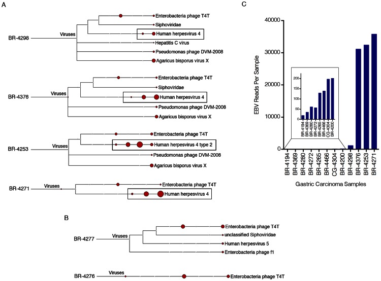
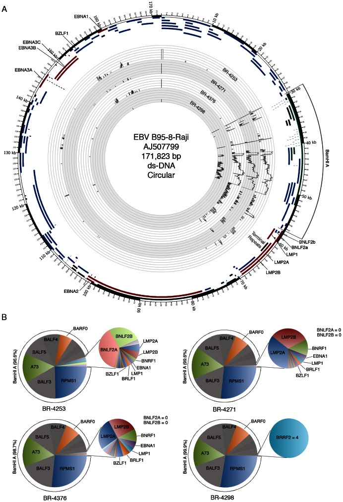
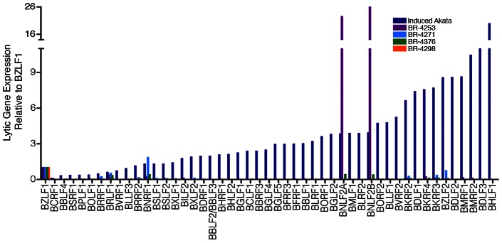
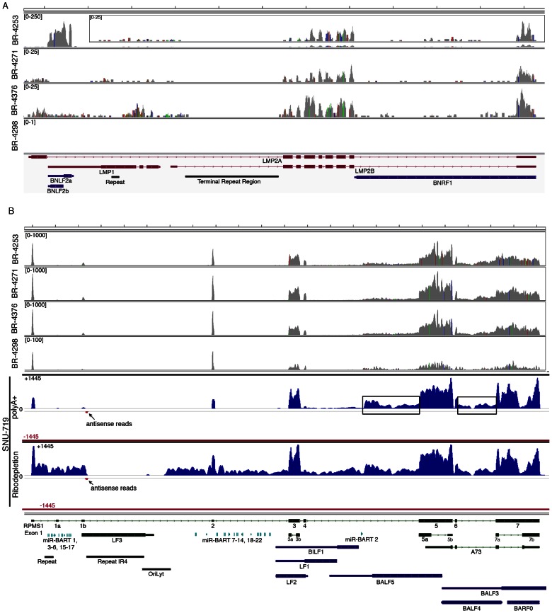
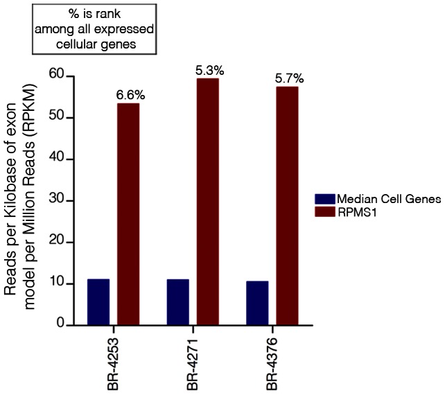
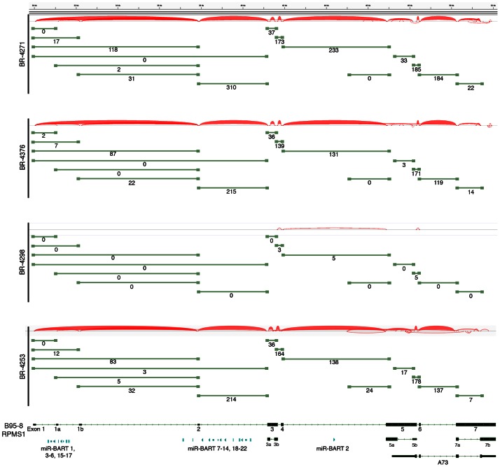
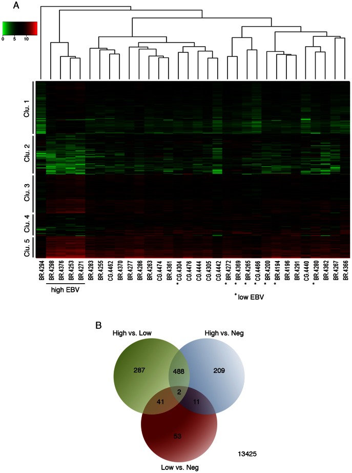
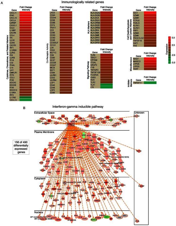
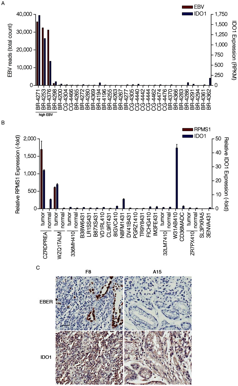
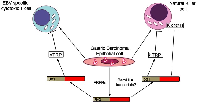
Epstein-Barr virus (EBV) is associated with roughly 10% of gastric carcinomas worldwide (EBVaGC). Although previous investigations provide a strong link between EBV and gastric carcinomas, these studies were performed using selected EBV gene probes. Using a cohort of gastric carcinoma RNA-seq data sets from The Cancer Genome Atlas (TCGA), we performed a quantitative and global assessment of EBV gene expression in gastric carcinomas and assessed EBV associated cellular pathway alterations. EBV transcripts were detected in 17% of samples but these samples varied significantly in EBV coverage depth. In four samples with the highest EBV coverage (hiEBVaGC - high EBV associated gastric carcinoma), transcripts from the BamHI A region comprised the majority of EBV reads. Expression of LMP2, and to a lesser extent, LMP1 were also observed as was evidence of abortive lytic replication. Analysis of cellular gene expression indicated significant immune cell infiltration and a predominant IFNG response in samples expressing high levels of EBV transcripts relative to samples expressing low or no EBV transcripts. Despite the apparent immune cell infiltration, high levels of the cytotoxic T-cell (CTL) and natural killer (NK) cell inhibitor, IDO1, was observed in the hiEBVaGCs samples suggesting an active tolerance inducing pathway in this subgroup. These results were confirmed in a separate cohort of 21 Vietnamese gastric carcinoma samples using qRT-PCR and on tissue samples using in situ hybridization and immunohistochemistry. Lastly, a panel of tumor suppressors and candidate oncogenes were expressed at lower levels in hiEBVaGC versus EBV-low and EBV-negative gastric cancers suggesting the direct regulation of tumor pathways by EBV.


Conflict of interest statement

The authors, Teresa A. Lehman and Michael B. Seddon are currently under the employment of BioServe Biotechnologies, Ltd. BioServe Biotechnologies, Ltd provided funds, resources and equipment to conduct some of the studies and provided salary and benefits for Teresa A. Lehman and Michael B. Seddon. This does not alter our adherence to all PLoS Pathogens policies on sharing data and materials.

Figures

Figure 1
Figure 1. Detection of EBV in gastric carcinoma samples.
Four gigabytes of deduplicated RNA-seq read data from each of the seventy-one gastric carcinoma samples were analyzed using RNA CoMPASS. The virome branch of the taxonomy trees for the four samples with the highest number of EBV reads (A) and two EBV-negative samples (B) were generated using the metagenome analysis tool, MEGAN 4. (C) For a more in depth analysis of EBV reads, the combined sequence read files for each sample were aligned to the EBV genome and the hg19 human genome assembly using the genome aligner, Novoalign. Of the EBV-positive samples, four samples were identified as having high numbers of EBV reads while eight were found to have low but detectable numbers of EBV reads (see Figure S2 for plot of EBV reads per 1,000,000 human mapped reads).
Figure 2
Figure 2. Genome wide analysis of EBV gene expression.
(A) An annotated Circos plot depicting EBV read coverage across the EBV genome. Coverage graphs display the number of reads mapping to each nucleotide position of the genome and are depicted in log scale. Expandable log and non-log plots are provided in Figures S3 and S4. Note that alignments were performed using a genome that was split between the BBLF2/3 and the BGLF3.5 lytic genes rather than at the terminal repeats to accommodate coverage of splice junctions for the latency membrane protein, LMP2. The natural termini of the linear genome, the terminal repeats, are shown in the lower right quadrant of the graph. Coverage data is plotted relative to the modified B95-8 genome containing Raji genome sequences (Genbank accession number AJ507799). Blue features represent lytic genes, red features represent latency genes, green features represent potential non-coding genes, aquamarine features represent microRNAs, and black features represent non-gene features (repeat regions and origins of replication, for example). (B) Pie charts displaying read counts across EBV gene features. Because the BNLF2a/b region is contained within the LMP1 gene, total LMP1 read counts were inferred by determining the counts within the unique LMP1 sequences, multiplying by the total length of LMP1, and dividing by the length of the unique region. BNLF2a/b counts were calculated by determining the number of reads within the BNLF2a/b locus and subtracting the inferred number of LMP1 reads derived from within the BNLF2a/b coordinates (i.e. number of LMP1 reads within the unique region times the length of the overlap region divided by the length of the unique region). Leftward oriented genes within the BamHI A region are shown in grey. This representation indicates uncertainty due to the finding of primarily rightward transcription across these genes in the gastric carcinoma cell line SNU-719 using directional sequencing methods (see below).
Figure 3
Figure 3. Abortive lytic gene expression.
EBV lytic gene expression in EBVaGC samples. Lytic gene expression relative to BZLF1 represents RPKMs (reads per kilobase of exon model per million mapped reads) for each indicated gene divided by the RPKMs of BZLF1 for the respective biological sample. For reference to a productive replication setting, samples were compared to the lytic gene expression profile in reactivated Akata cells.
Figure 4
Figure 4. EBV gene expression analysis.
Detailed read coverage data for the LMP2a, LMP1, and BNLF2a/b genes (A) and the RPMS1/BamHI A regions (B) of the EBV genome. Data was displayed using the Integrative Genomics Viewer (IGV) using the modified B95-8 genome containing Raji genome sequences (Genbank accession number AJ507799). The y axis represents the number of reads at each nucleotide position of the genome. Blue features represent lytic genes, red features represent latency genes, green features represent potential non-coding genes, aquamarine features represent microRNAs, and black features represent non-gene features (repeat regions and origins of replication, for example). In panel (A), coverage graphs for BR-4253 is scaled to a maximum read level of 250 reads (the BR-4253 inset displays the data with a max read level of 25), the BR-4271 and BR-4376 graphs are scaled to a max read level of 25, while the max read level for BR-4298 is 1. For coverage across the RPMS1/BamHI A region (B), BR-4253, BR-4271, and BR-4376 are scaled to 1,000 reads, while BR-4298 is scaled to 100. Strand specific sequencing from SNU-719 cells of the RPMS1/BamHI A region is also displayed. The top 2 tracks are from poly(A) selected RNA and the bottom 2 tracks are from Ribo-Zero depleted RNA. The read coverage for the sense strand is displayed in blue with positive values while the antisense strand is displayed in red with negative values. The scale is + or −1,445 reads for the sense and antisense strands.
Figure 5
Figure 5. EBV transcripts from RPMS1 are among the highest expressed genes in EBVaGCs.
RPKM values calculated using reads across all RPMS1 exons are shown with respect to the median expression of all expressed cellular genes (expressed genes defined as cellular genes with greater than 1 RPKM). The percentage values above each RPMS1 bar represents the rank of RPMS1 expression in the respective sample among all expressed cellular genes in that sample.
Figure 6
Figure 6. Alternative splicing in the EBV BamH1 A region in EBVaGCs.
RNA-seq data from BR-4271, BR-4376, and BR-4298 and BR-4253 was analyzed using the TopHat aligner to obtain splice junction information. Samples with the type I strain of EBV, BR-4271, BR-4376, BR-4298, and BR-4253 were aligned to the type I genome, B95-8/Raji (Genbank accession number AJ507799). Junctions were visualized using Integrative Genomic Viewer (IGV) . Thickness of red junction features correlates with the number of reads for the respective junction. The number of junction spanning reads for each junction is indicated below each olive green junction feature.
Figure 7
Figure 7. Cluster analysis of EBV-associated gastric carcinoma samples.
(A) A representative cohort of 32 gastric carcinoma samples (12 EBV-positive and 20 EBV-negative) were grouped using hierarchical clustering and are displayed with an expression heat map of the 490 genes that were found to be significantly differentially expressed in high EBV. (B) The cohort of 32 gastric carcinoma samples was divided into three categories (high EBV, low EBV, and negative). These categories were subjected to differential gene expression analysis using edgeR. The Venn diagram displays the numbers of all statistically significant differentially expressed genes. Statistical significance was determined by an adjusted P value<0.05.
Figure 8
Figure 8. High numbers of infiltrating immune cellular genes are detected in EBVaGC.
(A) Significant immunologically related genes differentially expressed in EBVaGC are represented in a heat map. The log2 fold change intensities are represented by the color gradient with red corresponding to the highest intensity and green corresponding to the lowest. (B) Interferon-gamma (IFNG) associated genes differentially expressed in EBVaGC are displayed in a diagram.
Figure 9
Figure 9. High levels of IDO1 in high EBV positive gastric carcinomas.
(A) Gene expression profile of the cohort of 32 gastric carcinoma samples (12 EBV-positive and 20 EBV-negative). Both total EBV reads and IDO1 expression (RPKM-reads per kilobase of exon model per million mapped reads) are represented as red and blue columns, respectively. (B) Gene expression profile of the cohort of 21 Vietnamese gastric carcinomas and 5 normal adjacent samples. Both relative RPMS1 expression (-fold) and relative IDO1 expression (-fold) are represented as red and blue columns and are the fold difference compared to the average of normal adjacent control values. (C) Images of paraffin-embedded human gastric carcinoma probed for EBER using in situ hybridization or IDO1 staining with immunohistochemistry. F8 and A15 each represent a specific gastric carcinoma on the tissue array selected to be closely matched with respect to age, tumor grade and stage. Scale bar represents 50 µm.
Figure 10
Figure 10. Model for EBV modulation of cytotoxic T-cell and natural killer cell function in tumor microenvironment.
EBV infected gastric carcinoma cells recruit cytolytic immune cells such as T-cell and natural killer cells via unclear mechanisms. In addition, these cells induce an increase in interferon-gamma (IFNG) via EBERs and possibly BamHI A transcripts. Increased IFNG results in increased IDO1 resulting in depleted tryptophan. Depleted tryptophan results in T-cell and natural killer cell inhibition.

References

    1. Burke A, Yen T, Shekitka K, Sobin L (1990) Lymphoepithelial carcinoma of the stomach with Epstein-Barr virus demonstrated by polymerase chain reaction. Mod Pathol 3: 377–380. - PubMed
    1. Shibata D, Weiss L (1992) Epstein-Barr virus-associated gastric adenocarcinoma. Am J pathol 140: 769–774. - PMC - PubMed
    1. Tokunaga M, Land C, Uemura Y, Tokudome T, Tanaka S, et al. (1993) Epstein-Barr virus in gastric carcinoma. Am J Pathol 143: 1250–1254. - PMC - PubMed
    1. Morewaya J, Koriyama C, Akiba S, Shan D, Itoh T, et al. (2004) Epstein-Barr virus-associated gastric carcinoma in Papua New Guinea. Oncol Rep 12: 1093–1098. - PubMed
    1. Moore RA, Warren R, Freeman JD, Gustavsen JA, Chenard C, et al. (2011) The Sensitivity of Massively Parallel Sequencing for Detecting Candidate Infectious Agents Associated with Human Tissue. PLoS ONE 6: e19838. - PMC - PubMed

Publication types

MeSH terms