. 2013 Apr 20;9(2):314-22.
doi: 10.5114/aoms.2012.31410. Epub 2012 Nov 6.

Adenovirus-mediated RAR-β over-expression enhances ATRA-induced neuronal differentiation of rat mesenchymal stem cells

Affiliations

Adenovirus-mediated RAR-β over-expression enhances ATRA-induced neuronal differentiation of rat mesenchymal stem cells

Yang Bi et al. Arch Med Sci. .

Abstract

Introduction: The retinoic acid (RA) signaling pathway plays important roles in neural development. All-trans retinoic acid (ATRA) activates the RA signal by regulating RAR-β in mesenchymal stem cell (MSC)-derived neuron cells. Here, we try to investigate whether RAR-β over-expression can affect neuronal differentiation of MSCs.

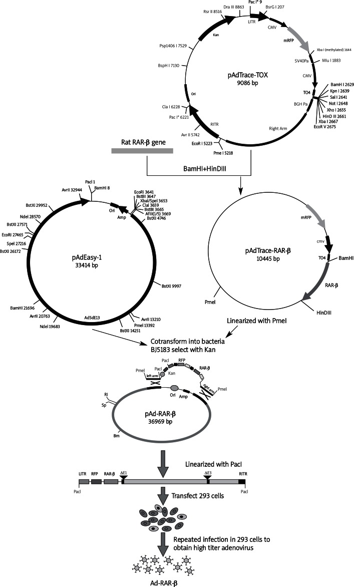
Material and methods: The RAR-β gene was constructed into adenovirus Ad-RAR-β by using the AdEasy system. The MSCs were infected with Ad-RAR-β. Real time-polymerase chain reaction (RT-PCR), Western blot and immunofluorescence were performed to detect the expression and localization of RAR-β. The MSCs were treated with 1 µmol/l ATRA and modified neuronal induction medium (MNM). Soma size and axon length of induced neurons were measured. Neural specific markers were detected by RT-PCR, western blot and immunofluorescence to evaluate neuronal differentiation.

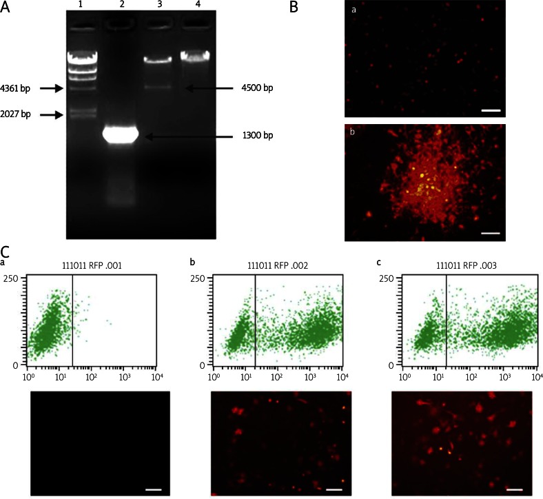
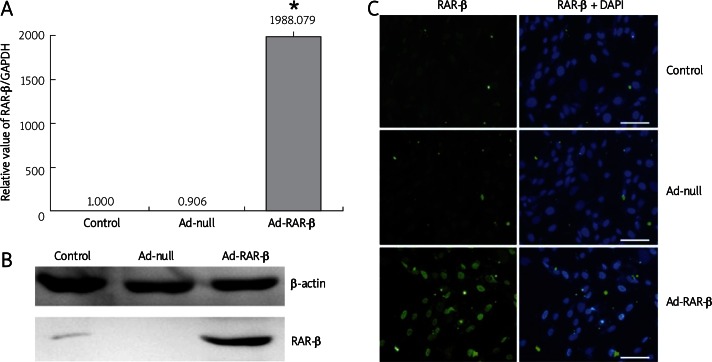
Results: The 1300 bp fragment of RAR-β gene was confirmed to be correctly cloned in the adenovirus vector. Cloudiness amplification of Ad-RAR-β was observed in HEK293 cells during package. After 48 h of Ad-RAR-β infection, about 70% of MSCs were RFP-positive. RAR-β expression was increased by about 1988-fold and located in the nucleus. RAR-β over-expression did not affect neuronal differentiation efficiency; however, soma size of induced neuron cells enlarged from 716.25 ±95.96 µm(2) to 1160.12 ±352.65 µm(2) and axon length from 64.17 ±11.88 µm to 83.98 ±13.69 µm. Neural markers other than nestin - NSE, MAP-2, Tau, and Tuj1 - were increased by 4- to 11-fold in RAR-β over-expressed neuron cells with ATRA/MNM induction compared with the Ad-null control group.

Conclusions: Our results have demonstrated that adenovirus-mediated RAR-β over-expression could facilitate neuron cell types of MSCs in vitro, indicating that the RAR-β-activated RA signal might be a vital factor in neuronal differentiation.

Keywords: all-trans retinoic acid; mesenchymal stem cells; neuronal differentiation; retinoic acid receptor β.


Figures

Figure 1
Figure 1
Construction and identification of adenovirus Ad-RAR-β. A – Identification of recombinant pAd-RAR-β plasmid by PCR amplification and enzyme digestion. 1 – λ-HinD III DNA marker, 2 – full length of RAR-β PCR amplified from pAd-RAR-β, 3 – pAd-RAR-β digested by Pac I enzyme, 4 – pAdEasy-1 bone vector digested by Pac I enzyme as negative control. B – Package of Ad-RAR-β in HEK293 cells. After digestion by Pac I enzyme and purification, pAd-RAR-β was liposome transfected into HEK293 cells. a – pAd-RAR-β transfected HEK293 cells at 1d. b – Cloudiness amplification of adenovirus Ad-RAR-β was observed in HEK293 cells at 10d pAd-RAR-β transfection. C – Infected efficacy of adenovirus Ad-RAR-β in MSCs. a – Uninfected MSCs, b – Ad-null infected MSCs, c – Ad-RAR-β infected MSCs (Scale bar = 200 µm)
Figure 2
Figure 2
Adenovirus Ad-RAR-β infection mediates over-expression of RAR-β in rat MSCs. The MSCs were infected with adenovirus Ad-RAR-β for 48 h, and untreated MSCs and Ad-null infected MSCs were used as controls. A – Total RNA of cells in different treated group was extracted and reverse transcribed into cDNA template. Expression of RAR-β was analyzed with GAPDH normalization. Real-time PCR results were confirmed in at least three batches of independent experiments (n = 3, * p < 0.05 vs. Ad-null/ ATRA/MNM group). B – Cells were lysed and subjected to SDS-PAGE and western blotting using RAR-β antibody. Equal loading of the samples was confirmed by β-actin expression. C – Localization and expression of RAR-β was detected by immunofluorescence staining. Cells were fixed and probed with primary antibodies against RAR-β, followed by staining with DyLight 488 labeled secondary antibodies and staining of nuclei with DAPI. Scale bar = 200 µm
Figure 3
Figure 3
Ad-RAR-β mediated RAR-β over-expression improves neuronal differentiation of rat MSCs. Cells were divided into 4 groups. Cells in control group were untreated, cells in ATRA/MNM induction group were induced with 1 µmol/l ATRA for 24 h, followed by MNM incubation for another 24 h. Before neuronal induction, cells in Ad-null/ATRA/MNM induction group and Ad-RAR-β/ATRA/MNM induction group were infected by adenovirus Ad-null and Ad-RAR-β for 48 h, respectively. A – Cell morphology, a – Control, b – ATRA/MNM treatment, c – Ad-null/ATRA/MNM treatment, d – Ad-RAR-β/ATRA/MNM treatment. B – Expression of nestin, NSE, MAP-2, and Tau was analyzed with GAPDH normalization (n = 3, * p < 0.05, Ad-RAR-β/ATRA/MNM vs. Ad-null/ATRA/MNM). Real-time PCR results were confirmed in at least three batches of independent experiments. C – Rat MSCs were treated with ATRA for 24 h and collected at the indicated time points with MNM culture, then lysed and subjected to SDS-PAGE and western blot using NSE and Tuj1 antibody. Equal loading of the samples was confirmed by β-actin expression. D – Immunofluorescence staining of neural specific markers. Cells were fixed and probed with antibodies against nestin, NSE, and Tuj1 followed by staining with DyLight 488 labeled secondary antibodies and staining of nuclei with DAPI. Scale bar = 200 µm
None
Illustration of construction of recombinant adenovirus vector containing rat RAR-β gene

References

    1. Martinez-Biarge M, Diez-Sebastian J, Kapellou O, et al. Predicting motor outcome and death in term hypoxic-ischemic encephalopathy. Neurology. 2011;76:2055–61. - PMC - PubMed
    1. Corti S, Nizzardo M, Nardini M, et al. Neural stem cell transplantation can ameliorate the phenotype of a mouse model of spinal muscular atrophy. J Clin Invest. 2008;118:3316–30. - PMC - PubMed
    1. Pimentel-Coelho PM, Mendez-Otero R. Cell therapy for neonatal hypoxic-ischemic encephalopathy. Stem Cells Dev. 2010;19:299–310. - PubMed
    1. Baksh D, Song L, Tuan RS. Adult mesenchymal stem cells: characterization, differentiation, and application in cell and gene therapy. J Cell Mol Med. 2004;8:301–16. - PMC - PubMed
    1. Hofstetter CP, Schwarz EJ, Hess D, et al. Marrow stromal cells form guiding strands in the injured spinal cord and promote recovery. Proc Natl Acad Sci U S A. 2002;99:2199–204. - PMC - PubMed

LinkOut - more resources