

doi: 10.1371/journal.pone.0063160.
Print 2013.
Differential distribution of the Ca (2+) regulator Pcp4 in the branchial arches is regulated by Hoxa2
Affiliations
- PMID: 23671666
- PMCID: PMC3650044
- DOI: 10.1371/journal.pone.0063160
Item in Clipboard
Differential distribution of the Ca (2+) regulator Pcp4 in the branchial arches is regulated by Hoxa2
PLoS One.
.
Abstract
Branchial arches are externally visible tissue bands in the head region of all vertebrate embryos. Although initially formed from similar components, each arch will give rise to different head and neck structures. In a screen designed to characterize the molecular control of branchial arch identity in mouse, we identified Pcp4 as a second branchial arch-specific molecular signature. We further show that the transcription factor Hoxa2 binds to Pcp4 chromatin and regulates Pcp4 expression in the second arch. Hoxa2 is also sufficient to induce Pcp4 expression in anterior first arch cells, which are Pcp4-negative.
Conflict of interest statement
Figures
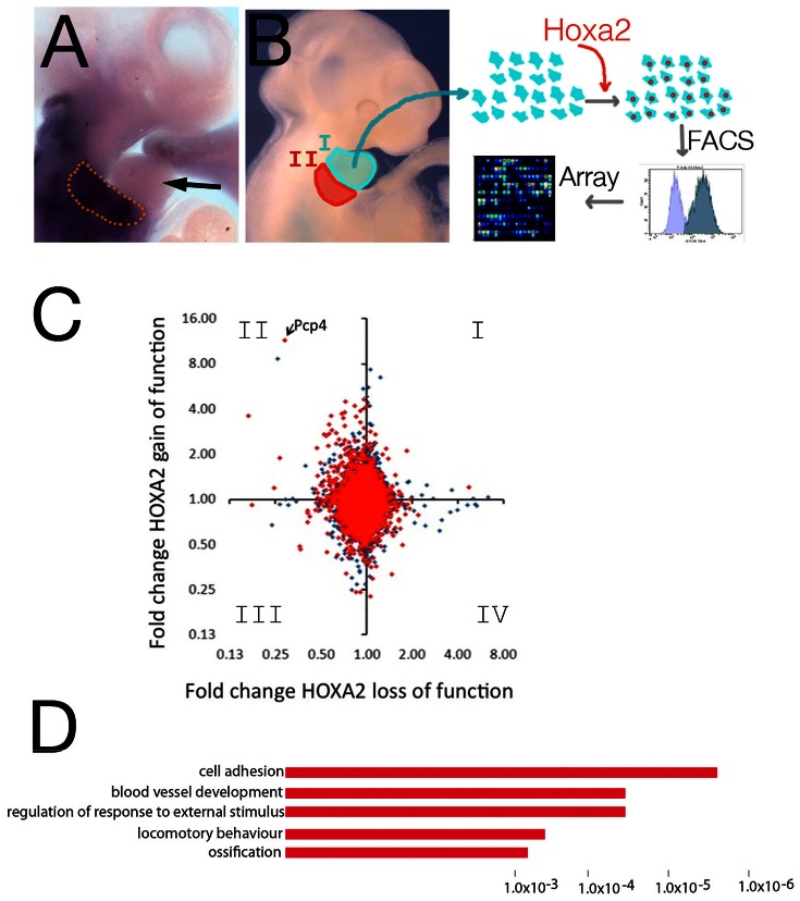
A, In situ hybridization using Hoxa2 probe shows Hoxa2 is mainly expressed in the IIBA (enclosed in the dotted line) and expression is excluded from the IBA (arrow). B, Schematic representation of the experiment: cells isolated from IBA are grown in vitro and transfected with Hoxa2-IRES-GFP (or GFP alone, control). RNA is extracted from GFP-positive cells and analyzed by microarray. C, Pairwise comparison of microarray experiments for Hoxa2 loss of function (x-axis) versus Hoxa2 gain of function (y-axis). Data are plotted as fold change against control in each case (axes in logarithmic scale base 2). Genes in red are nearby a Hoxa2-bound region in ChIP-seq (closest two genes to Hoxa2-bound region were included). D, Functional annotation of genes responsive to Hoxa2 gain of function only. The top over-represented categories are shown; the length of the bars corresponds to the P-values on the x-axis.
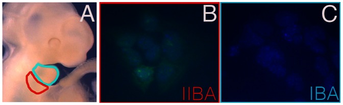
A, Head region of a midgestation mouse embryo, with first and second arch highlighted in turquoise and red, respectively. B,C. Immunofluorescence using Pcp4 antibody (green) stains cells isolated from IIBA (B), but not IBA (C). Nuclear staining is blue (DAPI).

A, High enrichment of the first intron of the Pcp4 gene compared to input (mm9, chr16∶96,717,332-96,718,404) in Hoxa2 ChIP-seq. Evolutionary Conserved Regions (ECRs) plot generated by the ECR Browser, comparing the genomic region bound by Hoxa2 between human, chimpanzee, and cow . B, Conventional ChIP on IIBA chromatin confirms enrichment of Hoxa2 to Pcp4. IP10 is a negative control gene. IgG is a non-specific negative control antibody.
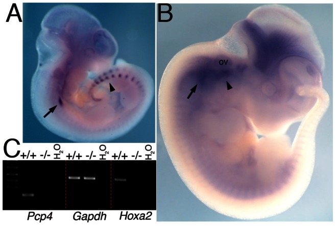
A,B. Whole mount ISH on E10.5 (A) and E11.5 (B) wild-type embryos, using Pcp4 probe. Pcp4 is expressed in the cranial ganglia (arrow, A-B) and dorsal root ganglia (arrowhead in A). One day later Pcp4 expression intensifies in the proximal area of the IIBA (arrowhead in B) in close proximity to the otic vesicle, and in the brain, and caudal expression disappears. C, Semiquantitative RT-PCR in wild type (+/+) and Hoxa2 mutant (−/−) E11.5 IIBA cDNA. Pcp4 is expressed in wild type and not Hoxa2 mutant IIBA cells. Gapdh is a positive control gene and Hoxa2 expression is confirmed in wild type and not Hoxa2 mutant IIBA cells. Ov, otic vesicle.
References
-
- Depew MJ, Lufkin T, Rubenstein JL (2002) Specification of jaw subdivisions by Dlx genes. Science, 298, 381–385. - PubMed
-
- Gendron-Maguire M, Mallo M, Zhang M, Gridley T (1993) Hoxa-2 mutant mice exhibit homeotic transformation of skeletal elements derived from cranial neural crest. Cell, 75, 1317–1331. - PubMed
-
- Rijli FM, Mark M, Lakkaraju S, Dierich A, Dolle P, Chambon P (1993) A homeotic transformation is generated in the rostral branchial region of the head by disruption of Hoxa-2, which acts as a selector gene. Cell, 75, 1333–1349. - PubMed
-
- Barrow JR, Capecchi MR (1999) Compensatory defects associated with mutations in Hoxa1 restore normal palatogenesis to Hoxa2 mutants. Development, 126, 5011–5026. - PubMed
-
- Manley NR, Capecchi MR (1997) Hox group 3 paralogous genes act synergistically in the formation of somitic and neural crest-derived structures. Dev Biol, 192, 274–288. - PubMed
Publication types
MeSH terms
Substances
Grants and funding
LinkOut - more resources
Full Text Sources
Other Literature Sources
Molecular Biology Databases
Miscellaneous
