. 2013 Apr 28;5(4):143-55.
doi: 10.4329/wjr.v5.i4.143.

Endovascular treatment of carotid cavernous sinus fistula: A systematic review

Affiliations

Endovascular treatment of carotid cavernous sinus fistula: A systematic review

Bora Korkmazer et al. World J Radiol. .

Abstract

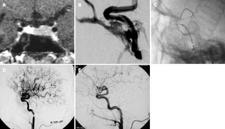
Carotid cavernous sinus fistulas are abnormal communications between the carotid system and the cavernous sinus. Several classification schemes have described carotid cavernous sinus fistulas according to etiology, hemodynamic features, or the angiographic arterial architecture. Increased pressure within the cavernous sinus appears to be the main factor in pathophysiology. The clinical features are related to size, exact location, and duration of the fistula, adequacy and route of venous drainage and the presence of arterial/venous collaterals. Noninvasive imaging (computed tomography, magnetic resonance, computed tomography angiography, magnetic resonance angiography, Doppler) is often used in the initial work-up of a possible carotid cavernous sinus fistulas. Cerebral angiography is the gold standard for the definitive diagnosis, classification, and planning of treatment for these lesions. The endovascular approach has evolved as the mainstay therapy for definitive treatment in situations including clinical emergencies. Conservative treatment, surgery and radiosurgery constitute other management options for these lesions.

Keywords: Carotid cavernous sinus fistula; Cavernous sinus; Endovascular treatment.


Figures

Figure 1
Figure 1
A thirty one-year-old man who was diagnosed with carotid cavernous sinus fistula which developed secondary to a motor vehicle accident. A: Digital subtraction angiogram view of left internal carotid artery (ICA) revealed laceration in the anterior loop and associated direct carotid cavernous sinus fistula; B: Balloon detachment failed because of the site and small calibre of the fistula orifice. Coil embolization of the fistula was then performed with preservation of the ICA; C: Post-procedural left ICA injection showed lack of residual filling, preservation of parent artery and detached coils at the site of fistula.
Figure 2
Figure 2
A fifty two-year-old woman who presented with galactorrhea due to hypophyseal adenoma underwent transsphenoidal surgery. During the operation internal carotid artery laceration and massive arterial hemorrhage occurred. A: T1 W C+ coronal magnetic resonance imaging view demonstrating hypointensity at the left hypophyseal region which was consistent with hypophyseal adenoma; B: Immediate defect source analysis revealed a defect at the anteromedial wall of right internal carotid artery (ICA) and associated carotid cavernous sinus fistula; C: Position of the stent-graft closing the orifice of the fistula; D: Postprocedural right ICA injection demonstrating complete obliteration of the fistula and concentric luminal stenosis at the petrous segment associated with mechanic vasospasm secondary to guide-wire and stent manipulation; E: Third month control angiography revealed regular parent artery contours, absence of recurrent fistula filling and intimal hyperplasia within the stent.
Figure 3
Figure 3
A thirty one-year-old male patient with right ophthalmoplegia following head trauma was found to have a direct carotid cavernous sinus fistula. A, B: Frontal (A) and lateral (B) digital subtraction angiogram views of right internal carotid artery (ICA) demonstrating laceration of ICA, pseudoaneurysm in the cavernous ICA and direct carotid cavernous sinus fistula; C: After considering the presence of the pseudoaneurysm, two detachable balloons were positioned to occlude the parent artery; D: Right CCA digital subtraction angiogram after balloon occlusion of the ICA showing complete obliteration of the fistula; E, F: Posttreatment left ICA (E) and left vertebral artery (F) injections demonstrating reconstruction of the right ICA area.
Figure 4
Figure 4
A forty-year-old woman with chemosis of the left eye and diplopia was found to have a dural carotid cavernous sinus fistula. A, B: Frontal (A) and lateral (B) injections of the right common carotid artery demonstrating left dural carotid cavernous sinus fistula with antegrade drainage; C: Access to the fistula site through the contralateral (right inferior petrous sinus) transvenous route and positioning of the microcatheter; D: Coil embolization within the microcather extending into the venous compartment of the fistula; E: Posttreatment frontal digital subtraction angiogram view of right internal carotid artery demonstrating obliteration of the fistula and lack of residual filling.
Figure 5
Figure 5
In extremely difficult cases of venous occlusion, stenosis, or marked tortuosity, access to the cavernous sinus can be provided by combined surgical and endovascular approaches. A, B: Left external carotid artery lateral (A) and left internal carotid artery lateral (B) digital subtraction angiogram views of an uncovered dural carotid cavernous sinus fistula which has antegrade drainage; C: Posterior access to the fistula site was not feasible, so access was gained through the superior ophthalmic vein following surgical angular vein cut-down; D: Coil detachment through a microcatheter into the fistula site; E: Posttreatment left common carotid artery injection revealed lack of residual filling of the fistula.

Similar articles

Cited by

References

    1. Barrow DL, Spector RH, Braun IF, Landman JA, Tindall SC, Tindall GT. Classification and treatment of spontaneous carotid-cavernous sinus fistulas. J Neurosurg. 1985;62:248–256. - PubMed
    1. Ernst RJ, Tomsick TA. Classification and angiography of carotid cavernous fistulas. In: Tomsick TA, editor. Carotid cavernous sinus fistula. Cincinnati: Digital Education Publishing; 1997. pp. 13–22.
    1. Coskun O, Hamon M, Catroux G, Gosme L, Courthéoux P, Théron J. Carotid-cavernous fistulas: diagnosis with spiral CT angiography. AJNR Am J Neuroradiol. 2000;21:712–716. - PMC - PubMed
    1. Ringer AJ, Salud L, Tomsick TA. Carotid cavernous fistulas: anatomy, classification, and treatment. Neurosurg Clin N Am. 2005;16:279–295. - PubMed
    1. Tjoumakaris SI, Jabbour PM, Rosenwasser RH. Neuroendovascular management of carotid cavernous fistulae. Neurosurg Clin N Am. 2009;20:447–452. - PubMed

LinkOut - more resources