

Meninges: from protective membrane to stem cell niche
- PMID: 23671802
- PMCID: PMC3636743
Meninges: from protective membrane to stem cell niche
Abstract
Meninges are a three tissue membrane primarily known as coverings of the brain. More in depth studies on meningeal function and ultrastructure have recently changed the view of meninges as a merely protective membrane. Accurate evaluation of the anatomical distribution in the CNS reveals that meninges largely penetrate inside the neural tissue. Meninges enter the CNS by projecting between structures, in the stroma of choroid plexus and form the perivascular space (Virchow-Robin) of every parenchymal vessel. Thus, meninges may modulate most of the physiological and pathological events of the CNS throughout the life. Meninges are present since the very early embryonic stages of cortical development and appear to be necessary for normal corticogenesis and brain structures formation. In adulthood meninges contribute to neural tissue homeostasis by secreting several trophic factors including FGF2 and SDF-1. Recently, for the first time, we have identified the presence of a stem cell population with neural differentiation potential in meninges. In addition, we and other groups have further described the presence in meninges of injury responsive neural precursors. In this review we will give a comprehensive view of meninges and their multiple roles in the context of a functional network with the neural tissue. We will highlight the current literature on the developmental feature of meninges and their role in cortical development. Moreover, we will elucidate the anatomical distribution of the meninges and their trophic properties in adult CNS. Finally, we will emphasize recent evidences suggesting the potential role of meninges as stem cell niche harbouring endogenous precursors that can be activated by injury and are able to contribute to CNS parenchymal reaction.
Keywords: Meninges; arachnoid mater; corticogenesis; leptomeninges; neural progenitors; neural stem cells; neurogenesis; pia mater; stem cell niche.
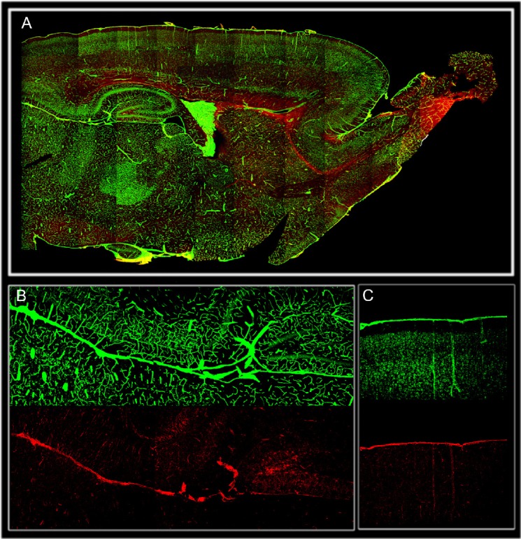
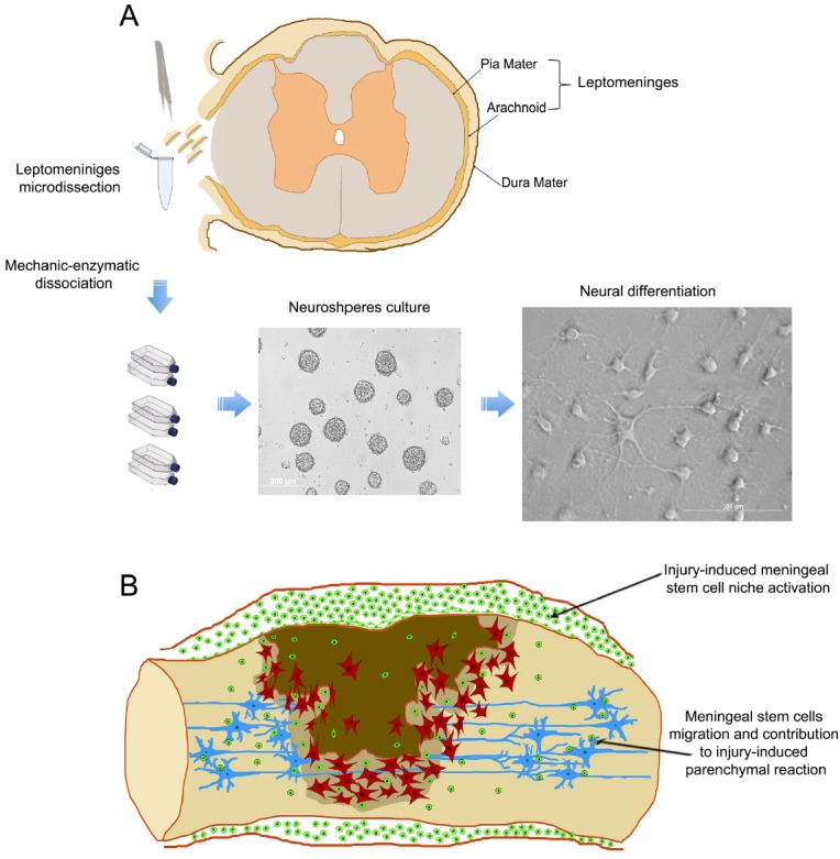
Figures
References
-
- Jacobson S, Marcus EM. Neuroanatomy for the Neuroscientist. Springer; 2008. pp. 325–331.
-
- Wilkins RH. Neurosurgical Classics. Thieme; 1992. p. 1.
-
- Beatriz M, Lopes S Meninges. Embryology. In: Joung H. Lee., editor. Meningiomas. Springer; 2009. pp. 25–29.
-
- Etchevers HC, Couly G, Vincent C, Le Douarin NM. Anterior cephalic neural crest is required for forebrain viability. Development. 1999;126:3533–3543. - PubMed
-
- O’Rahilly R, Müller F. The meninges in human development. J Neuropathol Exp Neurol. 1986;45:588–608. - PubMed
LinkOut - more resources
Full Text Sources
Other Literature Sources