. 2013 Sep;10(3):316-25.
doi: 10.1089/zeb.2013.0866. Epub 2013 May 14.

Deriving cell lines from zebrafish embryos and tumors

Affiliations

Deriving cell lines from zebrafish embryos and tumors

Suma Choorapoikayil et al. Zebrafish. 2013 Sep.

Abstract

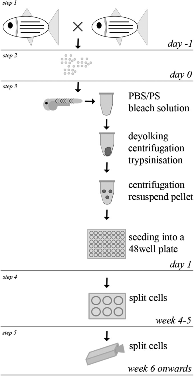
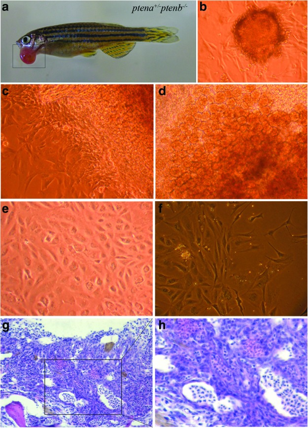
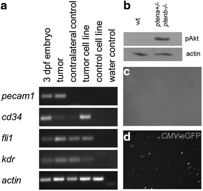
Over the last two decades the zebrafish has emerged as a powerful model organism in science. The experimental accessibility, the broad range of zebrafish mutants, and the highly conserved genetic and biochemical pathways between zebrafish and mammals lifted zebrafish to become one of the most attractive vertebrate models to study gene function and to model human diseases. Zebrafish cell lines are highly attractive to investigate cell biology and zebrafish cell lines complement the experimental tools that are available already. We established a straightforward method to culture cells from a single zebrafish embryo or a single tumor. Here we describe the generation of fibroblast-like cell lines from wild-type and ptenb(-/-) embryos and an endothelial-like cell line from a tumor of an adult ptena(+/-)ptenb(-/-) zebrafish. This protocol can easily be adapted to establish stable cell lines from any mutant or transgenic zebrafish line and the average time to obtain a pro-stable cell line is 3-5 months.


Figures

FIG. 1.
FIG. 1.
Workflow how to culture cells from an embryo. Schematical overview of single steps (1–5) is shown. Embryos are collected after natural mating (steps 1 and 2). Embryos are transferred to tubes and washed, bleached, deyolked, and trypsinized. Single-cell suspensions are transferred to a 48-well plate (step 3). After several weeks of culturing and monitoring, cells are split and transferred to a six-well plate (step 4). Confluent wells are split to a 25-cm2 flask (step 5).
FIG. 2.
FIG. 2.
Cells after seeding. (a) In some cases, parts of the embryo still remain after trypsinization (arrows); some single cells are indicated by arrowheads. (b) The embryo-cell suspension contains single floating cells. (c) After ∼1 h, first attached cells are visible. (d) Enlargement of indicated box in (c), showing an attached cell. (e) After 1 day, cells start to grow out of the embryo fragment (arrow). (f ) One day after culturing small colonies of cells are noticeable. Color images available online at www.liebertpub.com/zeb
FIG. 3.
FIG. 3.
Contamination and color switch of growth media. Small moving particles are indication of a contamination (arrow in a). A freshly added growth medium to a 75-cm2 flask (b, top flask) is shown. After one week, the color switched to deep pink (b, bottom flask). Color images available online at www.liebertpub.com/zeb
FIG. 4.
FIG. 4.
Culturing cells for about 5 weeks. (a) Four days after culturing, different cell types are detectable. (b–d) After 10 days, these cell types become more distinguishable. (e) Cells were passaged to a 6-well plate after 4 weeks of culturing in a 48-well plate and after reaching confluence, they were passaged further to 6-well plates. (f–h) After three passages in 3 weeks, single cell types are observed. Color images available online at www.liebertpub.com/zeb
FIG. 5.
FIG. 5.
Reculturing frozen cells. (a) One h after seeding, the first cells start to attach to the plate (arrows), single cells in suspension are indicated by arrowheads. (b) Cells adhere and grow after 2 days Color images available online at www.liebertpub.com/zeb.
FIG. 6.
FIG. 6.
Culturing cells from a tumor. (a) An external growing tumor mass (marked by box) in a ptena+/−ptenb−/− fish. Part of the tumor tissue was sliced and cut into several pieces. The rest of the tumor was fixed and embedded in paraffin. (b) After 7 days, cells have grown out of the tissue and form a cell layer surrounding the tissue. (c, d) After 3 weeks of culturing, different cell types are visible. (e) After 3–4 months, a single cell type is established (pro-cell line). (f ) After 1 year, a stable cell line is generated. (g, h) The fixed part of the tumor from panel (a), embedded in paraffin was sectioned transversally, and sections were stained with hematoxylin and eosin. (h) Enlargement of indicated box in (g), showing a hemangiosarcoma. Images were taken with 20× (g) and 40× (h), respectively. Color images available online at www.liebertpub.com/zeb
FIG. 7.
FIG. 7.
Zebrafish cells are amenable for experimental approaches. (a) Reverse transcription-PCR using RNA from 3 dpf embryos, hemangiosarcoma (tumor), contralateral control tissue, tumor cell line, a control cell line with the same genotype as the tumor-derived cell line (ptena+/−ptenb−/−) and H2O as a control. The primers that were used for the four endothelial markers and the loading control (actin beta 1, actb1) are depicted in Table 4. (b) Cells lacking Pten display an increased phosphoAkt level compared to wild type, illustrated by immunoblotting using phosphoAkt-specific antibodies. Anti-actin is included as a loading control. (c, d) Zebrafish cells are readily transfectable using lipofectamine (Invitrogen), illustrated by transfection of a CMV:eGFP construct that results in green fluorescent protein (GFP) expression throughout the cell. Pictures were taken using an AMG Evos microscope.

Similar articles

Cited by

References

    1. Lieschke GJ. Currie PD. Animal models of human disease: zebrafish swim into view. Nat Rev Genet. 2007;8:353–367. - PubMed
    1. Amatruda JF. Shepard JL. Stern HM. Zon LI. Zebrafish as a cancer model system. Cancer Cell. 2002;1:229–231. - PubMed
    1. Alders M. Hogan BM. Gjini E. Salehi F. Al-Gazali L. Hennekam EA, et al. Mutations in CCBE1 cause generalized lymph vessel dysplasia in humans. Nat Genet. 2009;41:1272–1274. - PubMed
    1. Cirstea IC. Kutsche K. Dvorsky R. Gremer L. Carta C. Horn D, et al. A restricted spectrum of NRAS mutations causes Noonan syndrome. Nat Genet. 42:27–29. - PMC - PubMed
    1. Jopling C. van Geemen D. den Hertog J. Shp2 knockdown and Noonan/LEOPARD mutant Shp2-induced gastrulation defects. PLoS Genet. 2007;3:e225. - PMC - PubMed

Publication types

LinkOut - more resources