. 2013 May 14:13:8.
doi: 10.1186/1472-6807-13-8.

A structural role for the PHP domain in E. coli DNA polymerase III

Affiliations

A structural role for the PHP domain in E. coli DNA polymerase III

Tiago Barros et al. BMC Struct Biol. .

Abstract

Background: In addition to the core catalytic machinery, bacterial replicative DNA polymerases contain a Polymerase and Histidinol Phosphatase (PHP) domain whose function is not entirely understood. The PHP domains of some bacterial replicases are active metal-dependent nucleases that may play a role in proofreading. In E. coli DNA polymerase III, however, the PHP domain has lost several metal-coordinating residues and is likely to be catalytically inactive.

Results: Genomic searches show that the loss of metal-coordinating residues in polymerase PHP domains is likely to have coevolved with the presence of a separate proofreading exonuclease that works with the polymerase. Although the E. coli Pol III PHP domain has lost metal-coordinating residues, the structure of the domain has been conserved to a remarkable degree when compared to that of metal-binding PHP domains. This is demonstrated by our ability to restore metal binding with only three point mutations, as confirmed by the metal-bound crystal structure of this mutant determined at 2.9 Å resolution. We also show that Pol III, a large multi-domain protein, unfolds cooperatively and that mutations in the degenerate metal-binding site of the PHP domain decrease the overall stability of Pol III and reduce its activity.

Conclusions: While the presence of a PHP domain in replicative bacterial polymerases is strictly conserved, its ability to coordinate metals and to perform proofreading exonuclease activity is not, suggesting additional non-enzymatic roles for the domain. Our results show that the PHP domain is a major structural element in Pol III and its integrity modulates both the stability and activity of the polymerase.


Figures
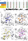
Figure 1
Figure 1
PHP domain metal-coordinating residues are not conserved. (A) Sequence alignment of C-family DNA polymerase PHP domains. The figure shows a selected set of sequences from our larger (47-sequence) alignment. Only sequences of polymerases that have been structurally or biochemically characterized were selected. For each polymerase the GI number and subtype within the C-family is indicated. For the conservation score diagram, the height of the bars is proportional to the conservation of the residues in our large alignment of C-family DNA polymerase sequences, as determined according to [17]. Black arrows at the top indicate the positions of variation in E. coli. (B) PHP domain cleft of C-family DNA polymerases. Metal-binding residues (or their substitutes in mutated PHP domains) are shown in ball and stick representation. Phosphate ions in E. coli Pol III and G. kaustophilus Pol C have been omitted for clarity.
Figure 2
Figure 2
A separate proofreading subunit coevolved with variant PHP domains. The trees were constructed using 50 selected sequences from our 47-sequence alignment of C-family DNA polymerases and 72 exonuclease sequences. Numbers indicate the GenInfo Identifier of the polymerase sequences. Two clades, corresponding to (1) α-, β- or γ-proteobacteria and (2) Thermus aquaticus and Aquifex aeolicus are shaded light orange and light grey in both trees, respectively. The tree in (B) shows whether the species to which the polymerase sequence corresponds contains an E. coli-like DNA polymerase III ϵ subunit homologue or not.
Figure 3
Figure 3
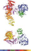
Structure of 3mPHP. The figure shows two orthogonal views of the 3mPHP structure determined at 2.9 Å resolution. The Pol III domains are coloured individually and the bound Zn2+ ions are shown as grey spheres.
Figure 4
Figure 4
Metal binding by the 3mPHP mutant. (A) Detail of the 3mPHP active site showing two peaks on the anomalous difference map contoured at 3.5 sigma shown in green. The two modelled Zn2+ ions are shown as spheres. Yellow dashed lines represent the distance (2.0 to 2.1 Å) between the side chains of the metal-binding residues and the centre of the two peaks. The (B) X-ray fluorescence scan of a zinc standard solution (grey) and of a 3mPHP protein sample (purple).
Figure 5
Figure 5
Restoration of metal-binding in E. coli Pol III does not induce exonuclease activity. (A) E. coli Pol III wild-type, 3mPHP and 4mPHP mutants show virtually no exonuclease activity in our measurements, as opposed to E. coli Pol III ϵ subunit that shows very robust activity under the same experimental conditions. The 5mPHP mutant shows some exonuclease activity, that is ~30-fold lower than that of the E. coli Pol III ϵ subunit. However, the metal-dependence of this activity is identical to that of the ϵ subunit. For both protein preparations, the exonuclease activity is stimulated by Mg2+ (B) and Mn2+, but is inhibited by Zn2+ (C; 0.3 mM MnCl2 background), suggesting that the observed activity for the 5mPHP preparation is due to contamination by ϵ subunit.
Figure 6
Figure 6
Mutations at the PHP domain decrease the overall stability of E. coli Pol III. The thermal and chemical stability of Pol III decreases gradually with the number of mutations introduced at the PHP domain, as measured by (A) temperature melt followed by circular dichroism or (B) through chemical denaturation using guanidine-hydrochloride titrations followed by circular dichroism and tryptophan fluorescence. Pol III shows apparent two-state unfolding.
Figure 7
Figure 7
Mutations at the PHP domain decrease Pol III polymerization activity. (A) Production of dsDNA was monitored by the intercalating dye PicoGreen. E. coli PHP mutants show substantially reduced polymerization activity. The decrease in activity correlates with the number of mutations introduced in the PHP domain. The relative polymerization rates of WT E. coli Pol III and the PHP mutants are shown in (B).

References

    1. Kornberg A, Baker TA. DNA Replication. 2. W. H: Freeman; 1992.
    1. Lamers MH, O’Donnell M. A consensus view of DNA binding by the C family of replicative DNA polymerases. P Natl Acad Sci Usa. 2008;105:20565–20566. doi: 10.1073/pnas.0811279106. - DOI - PMC - PubMed
    1. McHenry CS. DNA replicases from a bacterial perspective. Annu Rev Biochem. 2011;80:403–436. doi: 10.1146/annurev-biochem-061208-091655. - DOI - PubMed
    1. Huang YP, Ito J. DNA polymerase C of the thermophilic bacterium Thermus aquaticus: classification and phylogenetic analysis of the family C DNA polymerases. J Mol Evol. 1999;48:756–769. doi: 10.1007/PL00006520. - DOI - PubMed
    1. Lamers MH, Georgescu RE, Lee S-G, O’Donnell M, Kuriyan J. Crystal structure of the catalytic alpha subunit of E. coli replicative DNA polymerase III. Cell. 2006;126:881–892. doi: 10.1016/j.cell.2006.07.028. - DOI - PubMed

Publication types

Associated data