

doi: 10.1021/cn4000692.
Epub 2013 May 17.
Two-photon optical interrogation of individual dendritic spines with caged dopamine
- PMID: 23672485
- PMCID: PMC3750683
- DOI: 10.1021/cn4000692
Item in Clipboard
Two-photon optical interrogation of individual dendritic spines with caged dopamine
ACS Chem Neurosci.
.
Abstract
We introduce a novel caged dopamine compound (RuBi-Dopa) based on ruthenium photochemistry. RuBi-Dopa has a high uncaging efficiency and can be released with visible (blue-green) and IR light in a two-photon regime. We combine two-photon photorelease of RuBi-Dopa with two-photon calcium imaging for an optical imaging and manipulation of dendritic spines in living brain slices, demonstrating that spines can express functional dopamine receptors. This novel compound allows mapping of functional dopamine receptors in living brain tissue with exquisite spatial resolution.
Figures
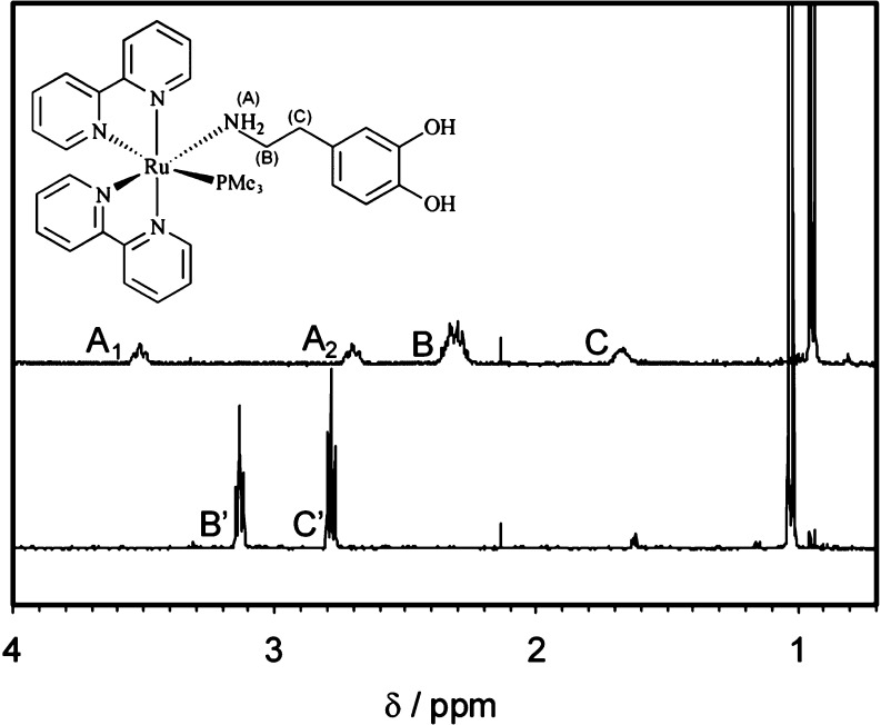
Structure (inset) and
aliphatic section of the 1H NMR
spectra of RuBi-Dopa in D2O before (top trace) and after
(bottom trace) irradiation into the NMR tube with a 525 nm green LED.
The doublets below 1.2 ppm belong to coordinated trimethylphosphine
(PMe3) in RuBi-Dopa (top trace) and in the aquo-complex
formed after photoreaction (bottom trace). Signals of coordinated
dopamine (B and C) are also apparent. The triplets A1 and
A2, corresponding to the coordinated −NH2, disappear after irradiation due to rapid exchange with D2O. The signals of the free dopamine methylenes (B′ and C′)
are indicative of the successful photorelease of the ligand.

UV–vis spectra of a 75 μM
aqueous solution of RuBi-Dopa
during photolysis at T = 25 °C and pH = 7. Samples
were irradiated at 405 nm, and spectra collected from 350 to 700 nm.
Inset: quantum yield calculation using eq 1.
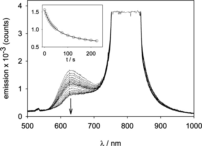
Photolysis of RuBi-Dopa in two-photon
regime. A solution containing
22 mM RuBi-Dopa was irradiated with an amplified Ti-Sa laser at 800
nm and the photoreaction followed through its emission band. Inset:
time dependence of the emission during photolysis. Details are given
in the text.
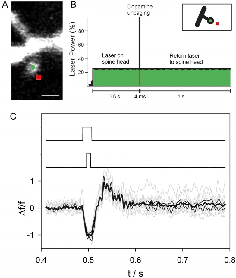
RuBi-Dopa
uncaging onto dendritic spines. (A) RuBi-Dopa is bath
applied to a cortical slice, and a layer five pyramidal neuron is
loaded with 200 μM Fluo-4 Ca2+ indicator and 200
μM Alexa-488. Scale bar = 1 μm. (B) Experimental configuration:
a Ti-Sapphire laser (820 nm) scans the spine head (at approximately
5–8 mW laser power on sample) first to monitor basal Ca2+ concentrations at the location marked with a circle in (A)
and then moves to a nearby location (square), where the laser power
is increased (to approximately 25 mW on the sample) to uncage dopamine
from RuBi-Dopa for 4 ms. After uncaging the laser goes back to initial
power and location on the spine head. (C) Spine fluorescent signals
(bottom traces; individual traces in gray (n = 10),
average trace in bold black, and ± StEr traces in black) showing
a sudden increase of Ca2+ concentration after the two-photon
uncaging of dopamine. The middle trace shows the uncaging pulse. The
fluorescent signal detection, by a photomultiplier tube (PMT), was
blocked for a few milliseconds before, during, and after the uncaging
pulse with the use of an ultrafast shutter (top trace) to prevent
potential PMT saturation and errors in signal detection.
References
-
- Schneier F. R.; Liebowitz M. R.; Abi-Dargham A.; Zea-Ponce Y.; Lin S. H.; Laruelle M. (2000) Low dopamine D(2) receptor binding potential in social phobia. Am. J. Psychiatry 157, 457. - PubMed
-
- Mink J. W. (2006) Neurobiology of basal ganglia and Tourette syndrome: basal ganglia circuits and thalamocortical outputs. Adv. Neurol. 99, 89–98. - PubMed
-
- Beaulieu J. M.; Gainetdinov R. R. (2011) The physiology, signaling, and pharmacology of dopamine receptors. Pharmacol. Rev 63, 182–217. - PubMed
-
- Adnet P.; Lestavel P.; Krivosic-Horber R. (2000) Neuroleptic malignant syndrome. Br. J. Anaesthesia 85, 129–135. - PubMed
Publication types
MeSH terms
Substances
Grants and funding
LinkOut - more resources
Full Text Sources
Other Literature Sources
