. 2013 Aug 30;394(1-2):49-54.
doi: 10.1016/j.jim.2013.05.002. Epub 2013 May 12.

Optimization of the ligature-induced periodontitis model in mice

Affiliations

Optimization of the ligature-induced periodontitis model in mice

Toshiharu Abe et al. J Immunol Methods. .

Abstract

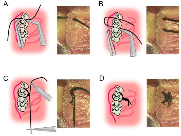
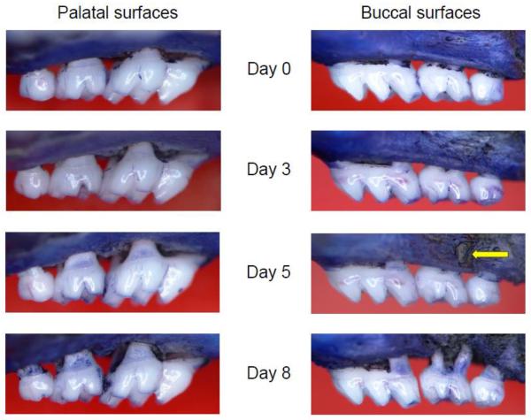
Periodontitis is a prevalent oral inflammatory disease that leads to alveolar bone loss and may exert an adverse impact on systemic health. Experimental animal models are critical tools to investigate mechanisms of periodontal pathogenesis and test new therapeutic approaches. The ligature-induced periodontitis model has been used frequently in relatively large animals, including non-human primates, to assess the host response and its effects on the tooth-supporting tissues (gingiva and bone) under well-controlled conditions. Although mice constitute the most convenient and versatile model for mechanistic immunological research (plethora of genetically engineered strains and immunological reagents), the tiny size of the murine oral cavity has presented technical challenges for ligature placement. In this report, we present a straightforward method for ligating the second maxillary molar tooth, and, moreover, identified the most appropriate sites for evaluating inflammatory bone loss in a valid and reproducible manner. These optimizations are expected to facilitate the use of the mouse ligature-induced periodontitis model and consequently contribute to better understanding of the immunopathological mechanisms of periodontitis.

Keywords: Bone loss; Inflammation; Ligature; Mouse model; Oral infection; Periodontitis.


Figures

Figure 1
Figure 1. Technical procedures of ligation
(A) 5–0 silk suture was passed through interdentium between second molar and third molar using Dumont forceps. (B) Suture was passed through interdentium between first molar and second molar using Dumont forceps. (C) Suture was looped around the second molar (taking care to remove the slack) using suture-tying forceps. (D) Suture was tied firmly using a triple-knot and excess suture was cut using spring scissors.
Figure 2
Figure 2. Quantification of anaerobic bacteria
Bacteria were extracted from recovered sutures (at the indicated days following placement of ligatures) and serial dilutions of bacterial suspensions were plated onto blood agar plates for anaerobic growth and CFU enumeration. Each symbol represents an individual mouse; small horizontal lines indicate the mean. *, P < 0.05, as compared with day 1.
Figure 3
Figure 3. Identification of sites susceptible to bone loss
(A) Schematics of sites subjected to measurements of CEJ-ABC distance. Key: M-P, mesio-palatal; M-B, mesio-buccal; P, palatal; B, buccal; D-P, disto-palatal; D-B, disto-buccal. (B) CEJ-ABC distance measurements at the palatal side, at the indicated days following placement of ligatures. (C) CEJ-ABC distance measurements at the buccal side, at the indicated days following placement of ligatures. *, P < 0.05 between day 8 and day 0.
Figure 4
Figure 4. Kinetics of total bone loss in palatal vs. buccal side
Groups of mice were euthanized 1, 3, 5, and 8 days after the placement of ligatures on the left maxillary molars. The 6-site total CEJ-ABC distance for the ligated side of each mouse was subtracted from the 6-site total CEJ-ABC distance of the contralateral unligated side of the same mouse. Panels A and B show measurements on the palatal and buccal surfaces, respectively. Data are means ± S.D. (n = 4) and negative values indicated bone loss relative to unligated baseline control. *, P < 0.05 compared to unligated baseline control.
Figure 5
Figure 5. Representative images of maxillae exhibiting time-dependent bone loss
The palatal side is contrasted with the buccal side in terms of time-dependent bone loss in ligature-induced periodontitis. Note the fenestration (arrow) forming on the buccal side of the second molar at day 5.

References

    1. Abe T, Hosur KB, Hajishengallis E, Reis ES, Ricklin D, Lambris JD, Hajishengallis G. Local complement-targeted intervention in periodontitis: proof-of-concept using a C5a receptor (CD88) antagonist. J Immunol. 2012;189:5442. - PMC - PubMed
    1. Abusleme L, Dupuy AK, Dutzan N, Silva N, Burleson JA, Strausbaugh LD, Gamonal J, Diaz PI. The subgingival microbiome in health and periodontitis and its relationship with community biomass and inflammation. ISME J Epub ahead of print. 2013 doi:10.1038/ismej.2012.174. - PMC - PubMed
    1. Assuma R, Oates T, Cochran D, Amar S, Graves DT. IL-1 and TNF antagonists inhibit the inflammatory response and bone loss in experimental periodontitis. J Immunol. 1998;160:403. - PubMed
    1. Bezerra MM, de Lima V, Alencar VB, Vieira IB, Brito GA, Ribeiro RA, Rocha FA. Selective cyclooxygenase-2 inhibition prevents alveolar bone loss in experimental periodontitis in rats. Journal of periodontology. 2000;71:1009. - PubMed
    1. Brecx MC, Nalbandian J, Ooya K, Kornman KS, Robertson PB. Morphological studies on periodontal disease in the cynomolgus monkey. II. Light microscopic observations on ligature-induced periodontitis. J Periodontal Res. 1985;20:165. - PubMed

Publication types