

Dyke-Davidoff-Masson syndrome: case report of fetal unilateral ventriculomegaly and hypoplastic left middle cerebral artery
- PMID: 23672850
- PMCID: PMC3666998
- DOI: 10.1186/1824-7288-39-32
Dyke-Davidoff-Masson syndrome: case report of fetal unilateral ventriculomegaly and hypoplastic left middle cerebral artery
Abstract
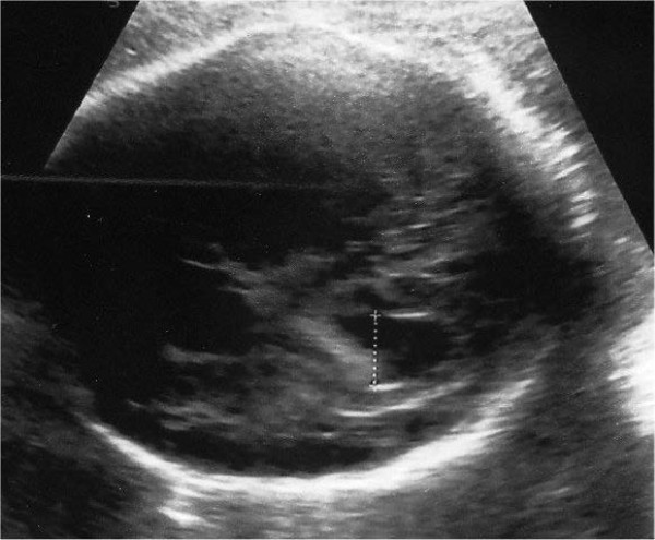
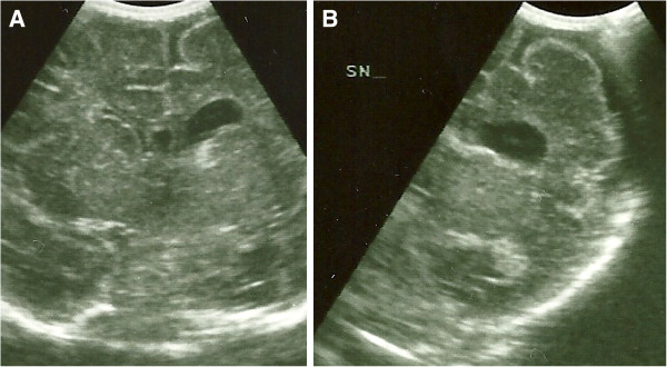
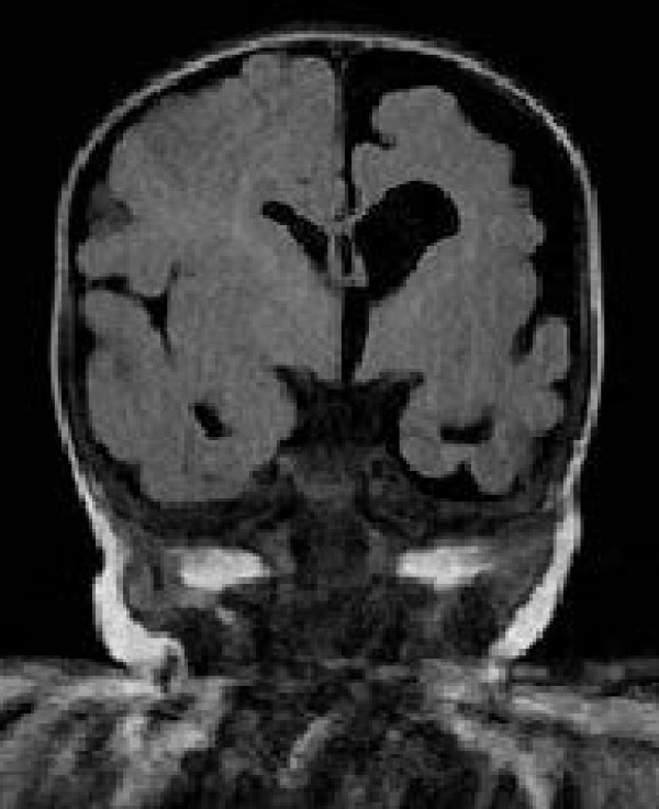
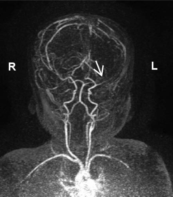
Prenatal ultrasonographic detection of unilateral cerebral ventriculomegaly arises suspicion of pathological condition related to cerebrospinal fluid flow obstruction or cerebral parenchimal pathology. Dyke-Davidoff-Masson syndrome is a rare condition characterized by cerebral hemiatrophy, calvarial thickening, skull and facial asymmetry, contralateral hemiparesis, cognitive impairment and seizures. Congenital and acquired types are recognized and have been described, mainly in late childhood, adolescence and adult ages. We describe a female infant with prenatal diagnosis of unilateral left ventriculomegaly in which early brain MRI and contrast enhanced-MRI angiography, showed cerebral left hemiatrophy associated with reduced caliber of the left middle cerebral artery revealing the characteristic findings of the Dyke-Davidoff-Masson syndrome. Prenatal imaging, cerebral vascular anomaly responsible for the cerebral hemiatrophy and the early clinical evolution have never been described before in such a young child and complete the acquired clinical descriptions in older children. Differential diagnosis, genetic investigations, neurophysiologic assessments, short term clinical and developmental follow up are described. Dyke-Davidoff-Masson syndrome must be ruled out in differential diagnosis of fetal unilateral ventriculomegaly. Early clinical assessment, differential diagnosis and cerebral imaging including cerebral MRI angiography allow the clinicians to diagnose also in early infancy this rare condition.
Figures
References
-
- Dyke CG, Davidoff LM, Masson CB. Cerebral hemiatrophy with homolateral hypertrophy of the skull and sinuses. Surg Gynecol Obstet. 1933;57:588–600.
-
- Pressler RM, Binnie C, Cooper R, Robinson R. Neonatal and paediatric clinical neurophisiology. 1. Amsterdam: Churchill Livingstone Elsevier; 2007. pp. 111–154.
Publication types
MeSH terms
LinkOut - more resources
Full Text Sources
Other Literature Sources
Medical
