

Subclinical macular findings in infants screened for retinopathy of prematurity with spectral-domain optical coherence tomography
- PMID: 23672969
- PMCID: PMC3737379
- DOI: 10.1016/j.ophtha.2013.01.028
Subclinical macular findings in infants screened for retinopathy of prematurity with spectral-domain optical coherence tomography
Abstract
Objective: To evaluate subclinical macular findings in premature patients at risk of retinopathy of prematurity (ROP) with the use of handheld spectral-domain optical coherence tomography (SD-OCT).
Design: Prospective, observational case series.
Participants: Forty-nine prematurely born neonates.
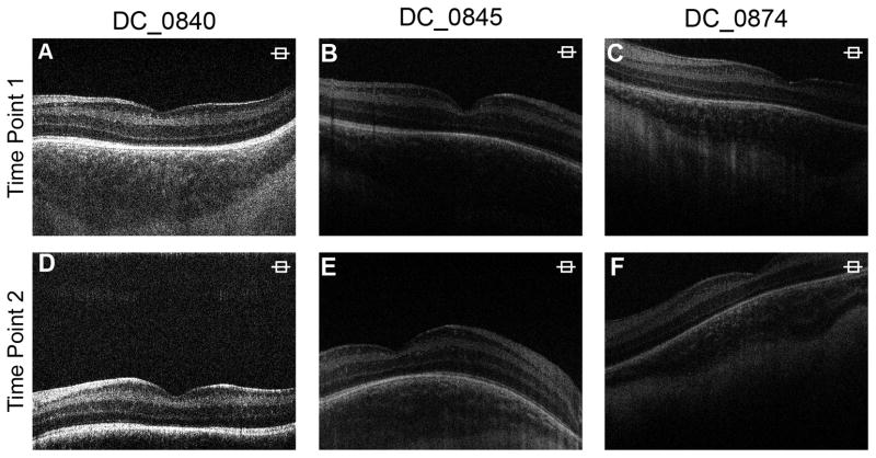
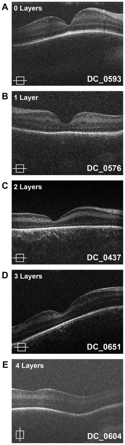
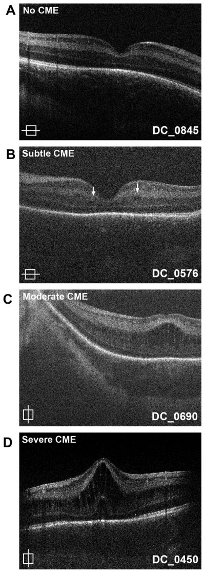
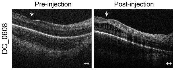
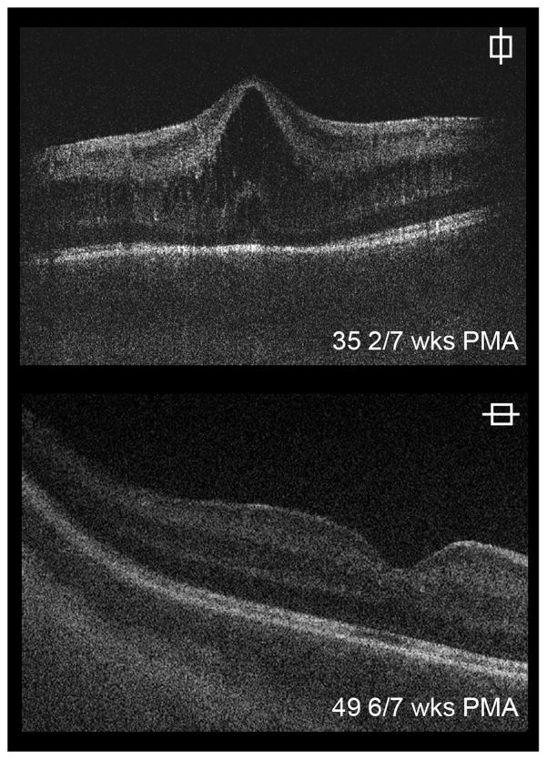
Methods: Forty-nine infants were imaged using a handheld SD-OCT. Images were acquired in nonsedated infants in the neonatal intensive care unit (NICU). Some patients were followed and reimaged over the course of several weeks. A total of 300 total images were acquired and evaluated for cystoid macular edema (CME) and persistence of inner retinal layers.
Main outcome measures: In vivo determination of foveal retinal lamination, image analysis, and clinical observation.
Results: A total of 241 (80%) of the images from 46 patients were usable (defined as having scans passing through the fovea with clearly identifiable retinal layers). Persistence of 1 or more inner retinal layers was seen in 43 of the patients with usable images (93%). Of the patients with at least 1 persistent layer, 17, 4, 8, 12, and 1, had a maximum ROP stage of 0, 1, 2, 3, and 4A, respectively. Cystoid macular edema was seen in 25 of the 46 patients (54%) during 1 or more imaging sessions. Cystoid macular edema was present in 9, 1, 5, 9, and 1 patient with maximum ROP stage of 0, 1, 2, 3, and 4A, respectively.
Conclusions: Our data suggest there is persistence of inner retinal layers in premature infants regardless of maximal ROP stage. Subclinical CME is seen in premature infants; however, CME does not appear to be correlated with ROP stage. This suggests that there may be other causes for the CME seen in this patient population. Hand-held SD-OCT imaging is a viable technique for evaluating subclinical macular findings in premature infants, although larger datasets are needed from multiple centers to further evaluate the generalizability of these findings.
Financial disclosure(s): The author(s) have no proprietary or commercial interest in any materials discussed in this article.
Copyright © 2013 American Academy of Ophthalmology. Published by Elsevier Inc. All rights reserved.
Figures
References
-
- Cryotherapy for Retinopathy of Prematurity Cooperative Group. Visual acuity at 10 years in Cryotherapy for Retinopathy of Prematurity (CRYO-ROP) study eyes: effect of retinal residua of retinopathy of prematurity. Arch Ophthalmol. 2006;124:199–202. - PubMed
-
- Akerblom H, Larsson E, Eriksson U, Holmström G. Central macular thickness is correlated with gestational age at birth in prematurely born children. Br J Ophthalmol. 2011;95:799–803. - PubMed
-
- Joshi MM, Trese MT, Capone A., Jr Optical coherence tomography findings in stage 4A retinopathy of prematurity: a theory for visual variability. Ophthalmology. 2006;113:657–60. - PubMed
-
- Ecsedy M, Szamosi A, Karko C, et al. A comparison of macular structure imaged by optical coherence tomography in preterm and full-term children. Invest Ophthalmol Vis Sci. 2007;48:5207–11. - PubMed
-
- Recchia FM, Recchia CC. Foveal dysplasia evident by optical coherence tomography in patients with a history of retinopathy of prematurity. Retina. 2007;27:1221–6. - PubMed
Publication types
MeSH terms
Grants and funding
LinkOut - more resources
Full Text Sources
Other Literature Sources
