. 2013 Jul 1;305(1):E119-31.
doi: 10.1152/ajpendo.00030.2013. Epub 2013 May 14.

Muscle-specific Pikfyve gene disruption causes glucose intolerance, insulin resistance, adiposity, and hyperinsulinemia but not muscle fiber-type switching

Affiliations

Muscle-specific Pikfyve gene disruption causes glucose intolerance, insulin resistance, adiposity, and hyperinsulinemia but not muscle fiber-type switching

Ognian C Ikonomov et al. Am J Physiol Endocrinol Metab. .

Abstract

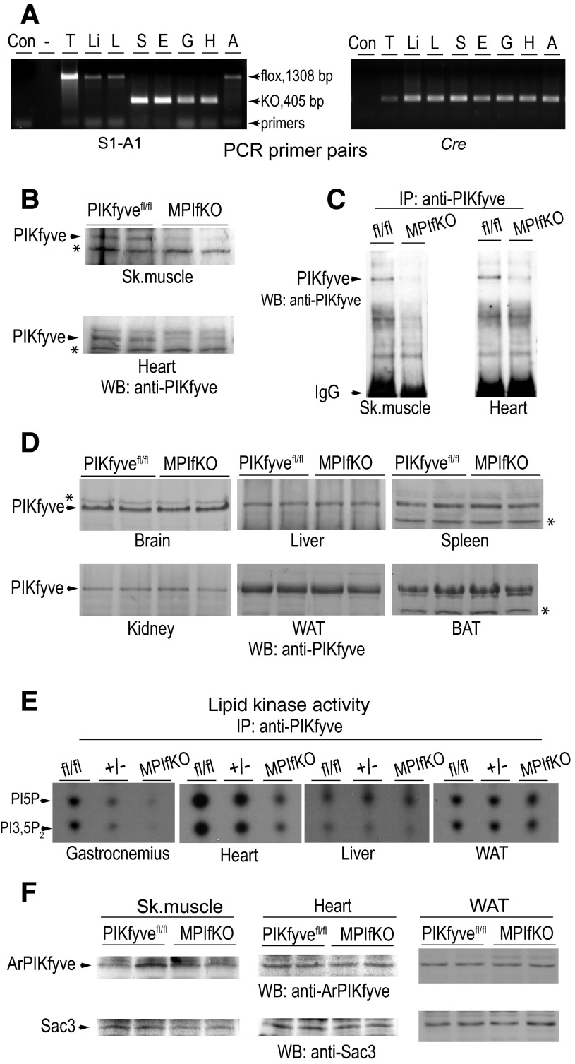
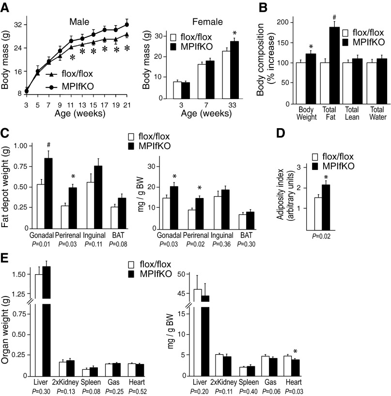
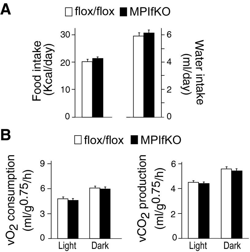
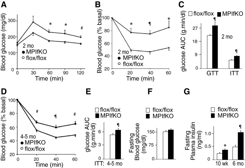
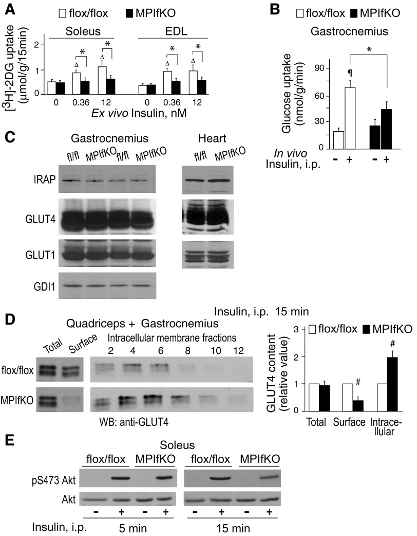
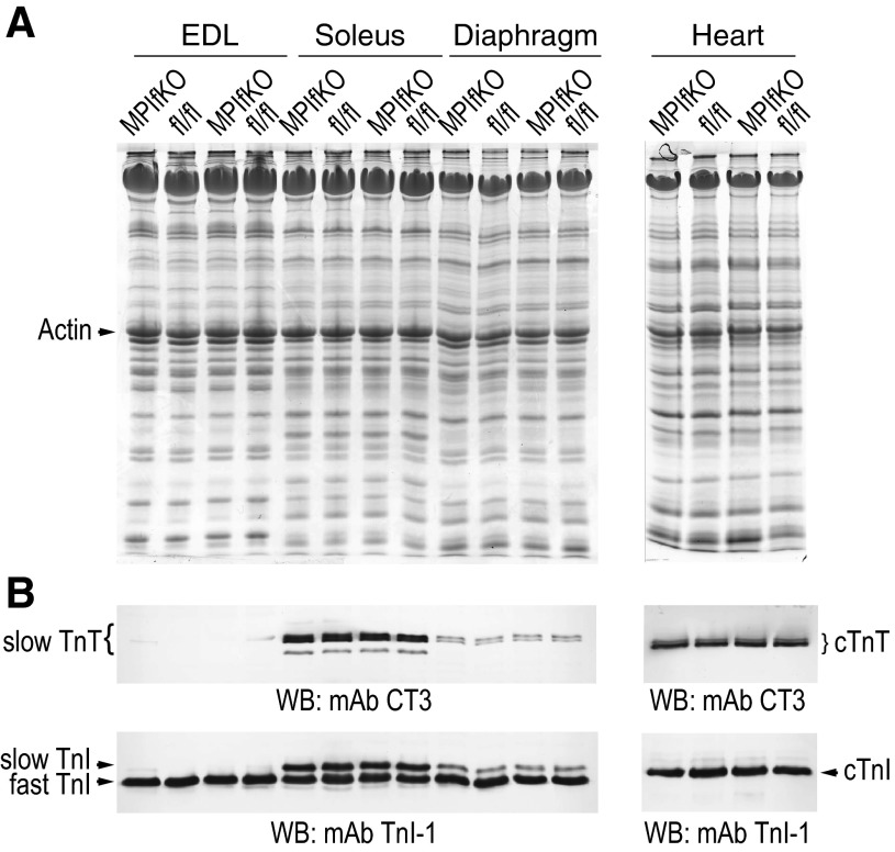
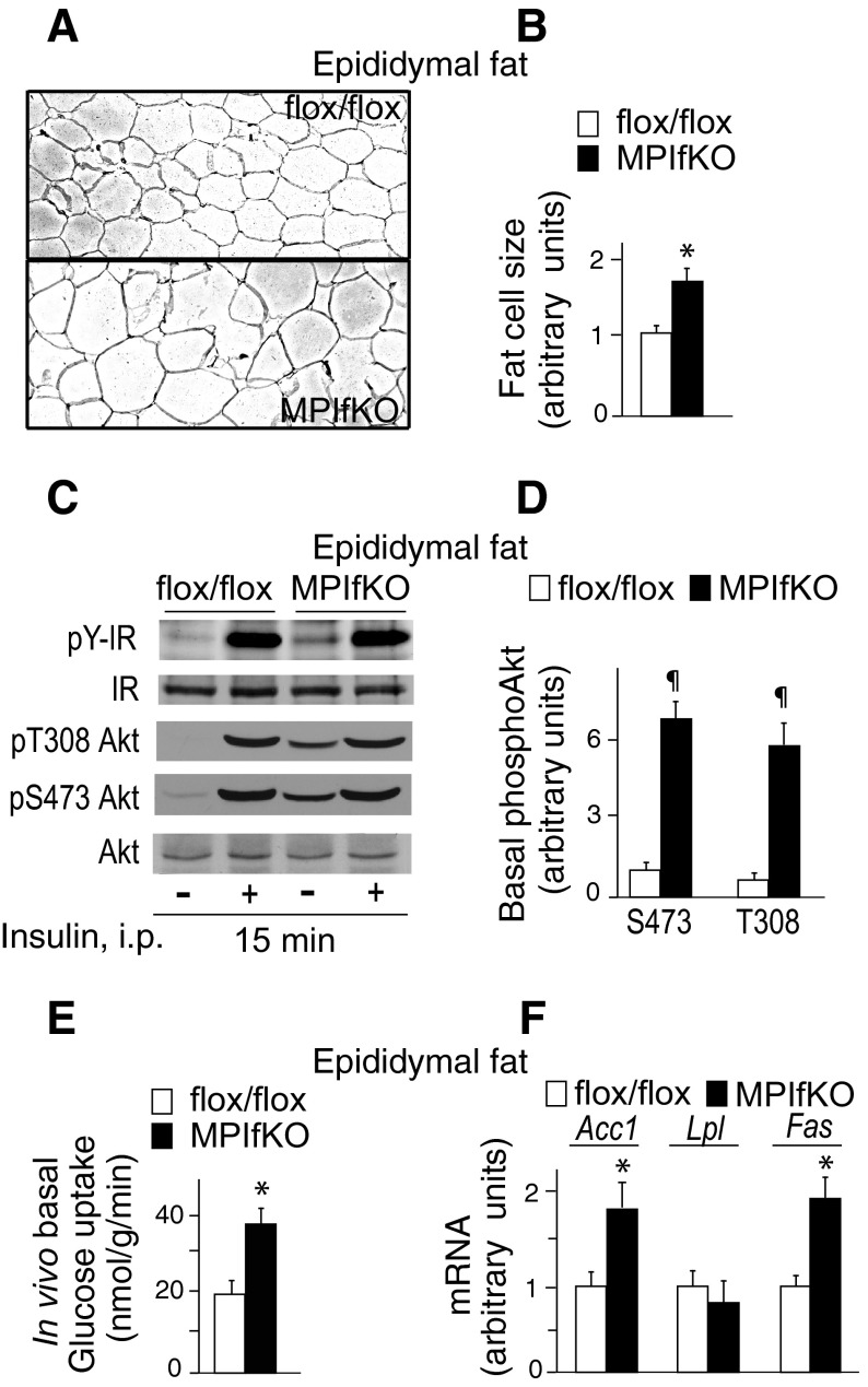
The evolutionarily conserved kinase PIKfyve that synthesizes PtdIns5P and PtdIns(3,5)P₂ has been implicated in insulin-regulated GLUT4 translocation/glucose entry in 3T3-L1 adipocytes. To decipher PIKfyve's role in muscle and systemic glucose metabolism, here we have developed a novel mouse model with Pikfyve gene disruption in striated muscle (MPIfKO). These mice exhibited systemic glucose intolerance and insulin resistance at an early age but had unaltered muscle mass or proportion of slow/fast-twitch muscle fibers. Insulin stimulation of in vivo or ex vivo glucose uptake and GLUT4 surface translocation was severely blunted in skeletal muscle. These changes were associated with premature attenuation of Akt phosphorylation in response to in vivo insulin, as tested in young mice. Starting at 10-11 wk of age, MPIfKO mice progressively accumulated greater body weight and fat mass. Despite increased adiposity, serum free fatty acid and triglyceride levels were normal until adulthood. Together with the undetectable lipid accumulation in liver, these data suggest that lipotoxicity and muscle fiber switching do not contribute to muscle insulin resistance in MPIfKO mice. Furthermore, the 80% increase in total fat mass resulted from increased fat cell size rather than altered fat cell number. The observed profound hyperinsulinemia combined with the documented increases in constitutive Akt activation, in vivo glucose uptake, and gene expression of key enzymes for fatty acid biosynthesis in MPIfKO fat tissue suggest that the latter is being sensitized for de novo lipid anabolism. Our data provide the first in vivo evidence that PIKfyve is essential for systemic glucose homeostasis and insulin-regulated glucose uptake/GLUT4 translocation in skeletal muscle.

Keywords: PIKfyve metabolic functions; PIKfyve muscle knockout; insulin resistance; insulin-regulated glucose transporter 4 translocation; muscle glucose uptake; prediabetes; systemic glucose homeostasis.


Figures

Fig. 1.
Fig. 1.
Validation of muscle-specific ablation of PIKfyve by PCR, Western blotting (WB), immunoprecipitation (IP), and in vitro kinase activity. A: representative genotyping with specific primer pairs for S1-A1 encompassing exon 6 and Cre, as indicated. A product of 405 bp is detected only in soleus (S), EDL (E), gastrocnemius (G), and heart (H) of the Cre-positive MPIfKO mice, whereas the other tissues, i.e., thymus (T), liver (Li), lung (L), and fat (A), make a product of 1,308 bp, having intact exon 6. B–D: RIPA+ lysates, derived from indicated tissues dissected from MPIfKO mice and PIKfyvefl/fl littermates, were clarified by centrifugation. Equal amounts of tissue protein (250 and 160 μg in muscle and other tissues, respectively) from each genotype were examined by WB with anti-PIKfyve antibodies (B and D) or by IP [2.5 mg, skeletal muscle (sk. muscle); and 875 μg, heart] with anti-PIKfyve prior to WB (C). Blots were reprobed with anti-GDI1 to normalize for loading (not shown). *Equal loading is also apparent by the identical intensity of the unspecific bands in the tissue samples. Shown are chemiluminescence detections from representative experiments with 2 mice/genotype for each condition out of 3–5 independent determinations. E: clarified fresh RIPA+ lysates derived from the indicated tissues dissected from MPIfKO and PIKfyvefl/fl mice underwent IP with anti-PIKfyve antibodies. Washed IPs were subjected to in vitro lipid kinase activity assay. Shown are representative autoradiograms of a TLC plate with resolved radiolabeled lipids, indicating that only in muscle of MPIfKO mice were the PIKfyve lipid products abrogated. In heterozygotes (+/−, genotype fl/WT; Cre+), these products were detected in gastrocnemius at significantly lower levels than in control (fl/fl;Cre−). F: clarified fresh RIPA+ lysates (250 μg protein) derived from the indicated tissues dissected from control and mutant mice were subjected to WB with antibodies against the partner proteins ArPIKfyve and Sac3. No significant changes between control and MPIfKO mice are apparent. Shown are chemiluminescence detections from representative experiments, with 2 mice/genotype for each condition out of 3–5 independent determinations. WAT, white adipose tissue; BAT, brown adipose tissue.
Fig. 2.
Fig. 2.
Increased body weight gain and adiposity in MPIfKO mice. A: rate of body weight gain in male (n = 7/group) and female mice (n = 4/group) fed a regular diet. B: in vivo body composition by EcoMRI (6-mo-old male mice; n = 7/group). CE: absolute or relative organ weights normalized per body weight and the adiposity index in 9- to 10-mo-old male mice (n = 8–10/group); #P < 0.01 and *P < 0.05 vs. control. Adiposity index was calculated by the following formula: total fat pad weight/(body weight − total fat pad weight).
Fig. 3.
Fig. 3.
Food/water intake and energy expenditure in MPIfKO mice. A: food/water consumption was measured in 9-wk-old male mice fed a regular diet over a period of 7 days. Note that the body weight at this age was similar between the mutant and control groups (n = 7/group). B: O2 consumption and CO2 production were measured in 10-wk-old male mice over a period of 72 h, subsequent to 48-h acclimation, and quantified separately for the light and dark cycles. The 2 groups show no differences (n = 7/group).
Fig. 4.
Fig. 4.
Systemic glucose intolerance and insulin resistance in MPIfKO mice. A and B: glucose levels were measured at indicated time points before and after 1.5 g/kg of glucose (A) or 0.75 U/kg of insulin (B), which were administered ip in 8- to 10-wk-old male mice; n = 6–8/group. C: the area under the blood glucose curves (AUC) during glucose (GTT) and insulin tolerance tests (ITT). D and E: glucose levels measured at indicated time points before and after ip administration of insulin (0.75 U/kg) in 4- to 5-mo-old male mice (n = 9–13/group; D) and AUC during ITT (E), indicating that insulin resistance persisted in older MPIfKO mice. F: fasting blood glucose concentration in 6-mo-old male mice; n = 9–13/group. G: fasting plasma insulin in 10-wk- (n = 6/group) and 6-mo-old mice (n = 7/group). *P < 0.05, #P < 0.01, and ¶P < 0.001 vs. controls.
Fig. 5.
Fig. 5.
Impaired insulin-dependent glucose uptake and glucose transporter (GLUT)4 translocation in muscle of MPIfKO. A: ex vivo glucose uptake was determined in soleus or extensor digitorum longus (EDL) muscles, excised from 10-wk-old mice, and incubated with insulin at the indicated concentrations for 15 min. 2-[3H]deoxyglucose ([3H]2DG) uptake was determined as described in materials and methods; n = 4–7/condition; ΔP < 0.05 vs. 0 insulin; *P < 0.05 vs. control mice. B: in vivo glucose uptake was measured in 14-h-starved 12-wk-old male mice that were administered [3H]2DG and l-[14C]glucose ip with or without insulin (1 U/kg), as described in materials and methods; n = 5/condition. ¶P < 0.001 vs. nontreated control group; *P < 0.05 between the insulin-treated groups. C: clarified fresh RIPA+ lysates isolated from gastrocnemius or cardiac muscle were subjected to immunoblotting with the indicated antibodies. Blots were reprobed with anti-GDI1 to normalize for loading. Shown are chemiluminescence detections from a representative experiment, with 2 mice/genotype out of 3–6 independent determinations. Apparent are the similar levels of GLUT4, insulin-responsive aminopeptidase (IRAP), and GLUT1 between MPIfKO and control mice. D: insulin (5 U/kg) was administered ip in 14-h-fasted mice. Fifteen minutes following injection, quadriceps (2×) and gastrocnemius (1×) were dissected and subjected to differential/sucrose velocity centrifugation, as detailed in materials and methods. Shown are chemiluminescence detections of an anti-GLUT4 blot from a representative experiment and quantitation by densitometry from 3 experiments (#P < 0.01). E: insulin (5 U/kg) was administered ip in 14-h-starved mice for 5 or 15 min. Soleus muscle was isolated and subjected to Western blotting analysis (65 μg of protein) with antibodies for phospho-Ser473 Akt or total Akt. Reduced Akt phosphorylation in MPIfKO muscle is evident at 15-min but not 5-min insulin. Shown are chemiluminescence detections from a representative experiment for each time point out of 3 independent determinations.
Fig. 6.
Fig. 6.
Unaltered muscle fiber type in MPIfKO mice despite metabolic abnormalities. A and B: protein extracts, prepared by homogenization in sample buffer of fresh muscle tissues from MPIfKO and control female mice (8 wk old), were subjected to SDS-PAGE in duplicate gels. Gels were either stained with Coomassie Blue R250 (A) or processed for immunoblotting with monoclonal antibodies (mAb) troponin I-1 (TnI-1) against muscle-specific myofilament protein troponin I (TnI), recognizing skeletal muscle fast/slow TnI and the cardiac muscle (c) TnI, and with mAb CT3 against muscle-specific troponin T (TnT), recognizing skeletal muscle slow TnT and the cardiac muscle TnT (B). Shown are Coomassie blue (A) and chemical detections (B) from a representative experiment. No changes were found between the control and MPIfKO mice in any of the muscles in 3 independent experiments.
Fig. 7.
Fig. 7.
Hypertophy, augmented basal Akt phosphorylation, and in vivo glucose uptake in MPIfKO fat. A and B: hematoxylin and eosin staining of paraffin-embedded epididymal fat tissue sections (A) showing profound hypertrophy in MPIfKO fat, quantified in B; n = 3/genotype; *P < 0.05 vs. control mice. C and D: constitutive Akt phosphorylation in MPIfKO fat. Insulin (5 U/kg) was administered ip in 14-h-starved mice. Fifteen minutes postinjection, epididymal fat tissue was isolated and subjected to Western blotting analysis (65 μg of protein) with antibodies for phospho-Thr308 Akt or phospho-Ser473 Akt, phosphotyrosine (P-Y), Akt, and insulin receptor (IR). Shown are chemiluminescence detections from a representative experiment (C) and quantitation of increased basal Akt phosphorylation in MPIfKO fat relative to Ser473 phosphorylation in controls (D); n = 5/group. ¶P < 0.001 vs. control mice. E: in vivo glucose uptake in epididymal fat was measured in 14-h-starved mice that were injected ip with [3H]2DG and l-[14C]glucose, as described in materials and methods; n = 5/genotype. *P < 0.05 vs. control mice. F: gene expression in epididymal fat from 12-wk-old mice; n = 6/genotype. *P < 0.05 vs. control mice. Acc1, acetyl-CoA carboxylase1; Lpl, lipoprotein lipase; Fas, fatty acid synthase.

References

    1. Abdul-Ghani MA, DeFronzo RA. Pathophysiology of prediabetes. Curr Diab Rep 9: 193–199, 2009 - PubMed
    1. Biddinger SB, Kahn CR. From mice to men: insights into the insulin resistance syndromes. Annu Rev Physiol 68: 123–158, 2006 - PubMed
    1. Björnholm M, Zierath JR. Insulin signal transduction in human skeletal muscle: identifying the defects in Type II diabetes. Biochem Soc Trans 33: 354–357, 2005 - PubMed
    1. Bridges D, Ma JT, Park S, Inoki K, Weisman LS, Saltiel AR. Phosphatidylinositol 3,5-bisphosphate plays a role in the activation and subcellular localization of mechanistic target of rapamycin 1. Mol Biol Cell 23: 2955–2962, 2012 - PMC - PubMed
    1. Bruning JC, Michael MD, Winnay JN, Hayashi T, Horsch D, Accili D, Goodyear LJ, Kahn CR. A muscle-specific insulin receptor knockout exhibits features of the metabolic syndrome of NIDDM without altering glucose tolerance. Mol Cell 2: 559–569, 1998 - PubMed

Publication types

MeSH terms