. 2013 Jul 1;305(1):E149-59.
doi: 10.1152/ajpendo.00040.2013. Epub 2013 May 14.

Adaptive β-cell proliferation increases early in high-fat feeding in mice, concurrent with metabolic changes, with induction of islet cyclin D2 expression

Affiliations

Adaptive β-cell proliferation increases early in high-fat feeding in mice, concurrent with metabolic changes, with induction of islet cyclin D2 expression

Rachel E Stamateris et al. Am J Physiol Endocrinol Metab. .

Erratum in

Abstract

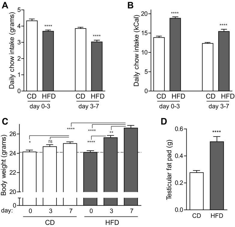
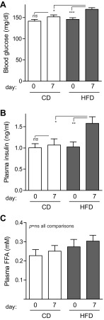
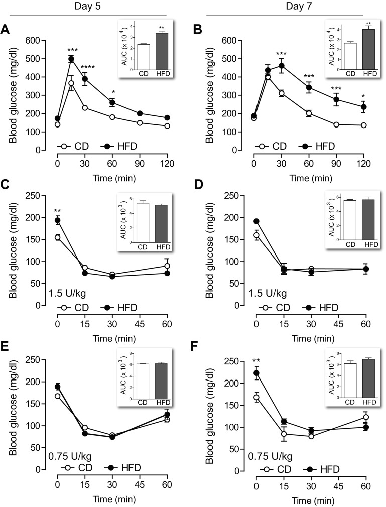
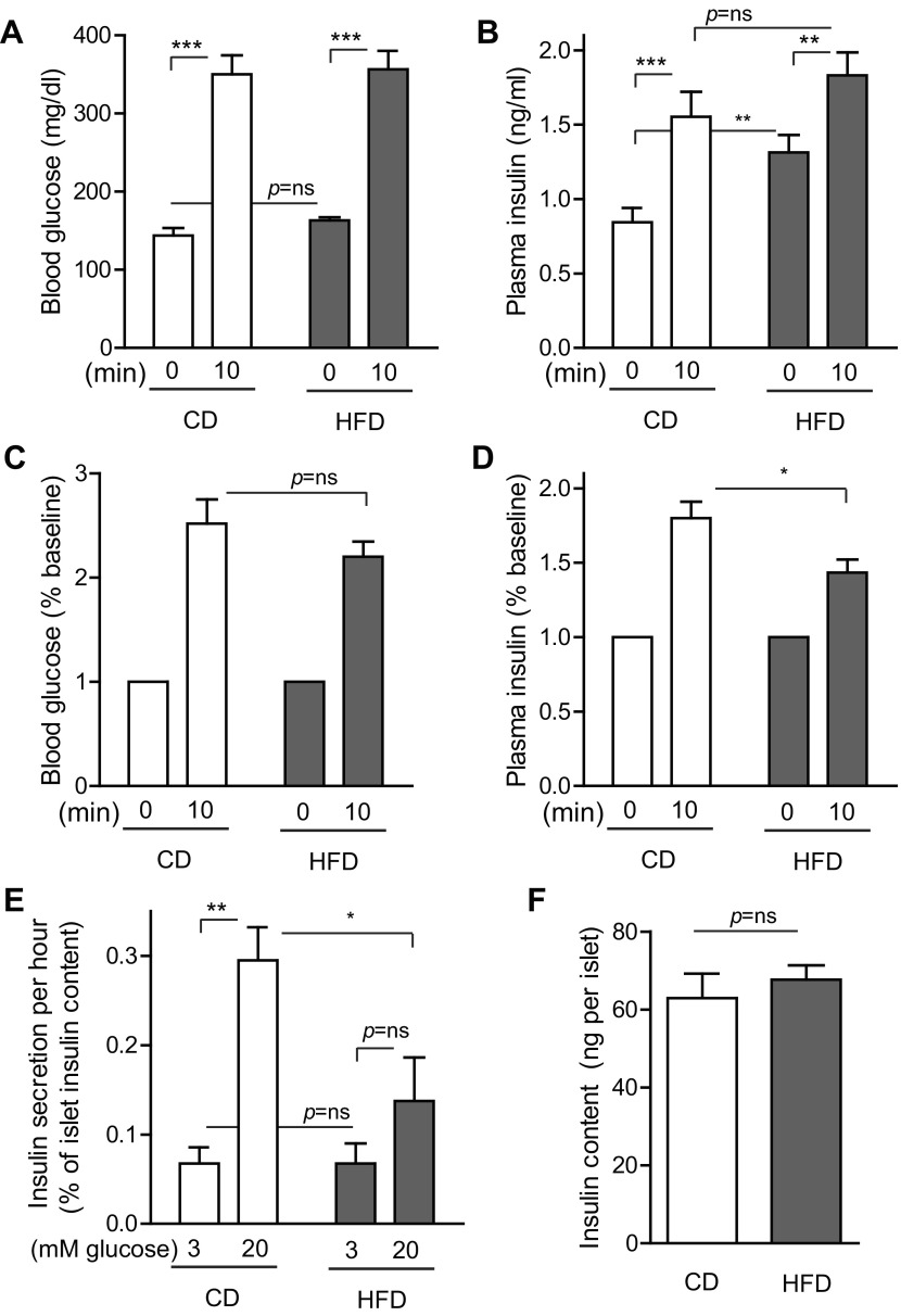
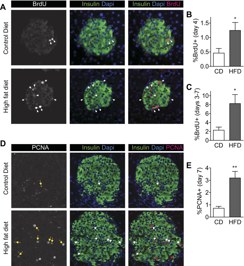
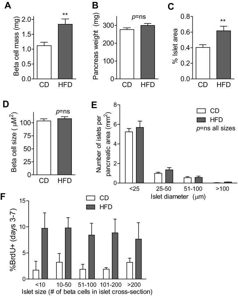
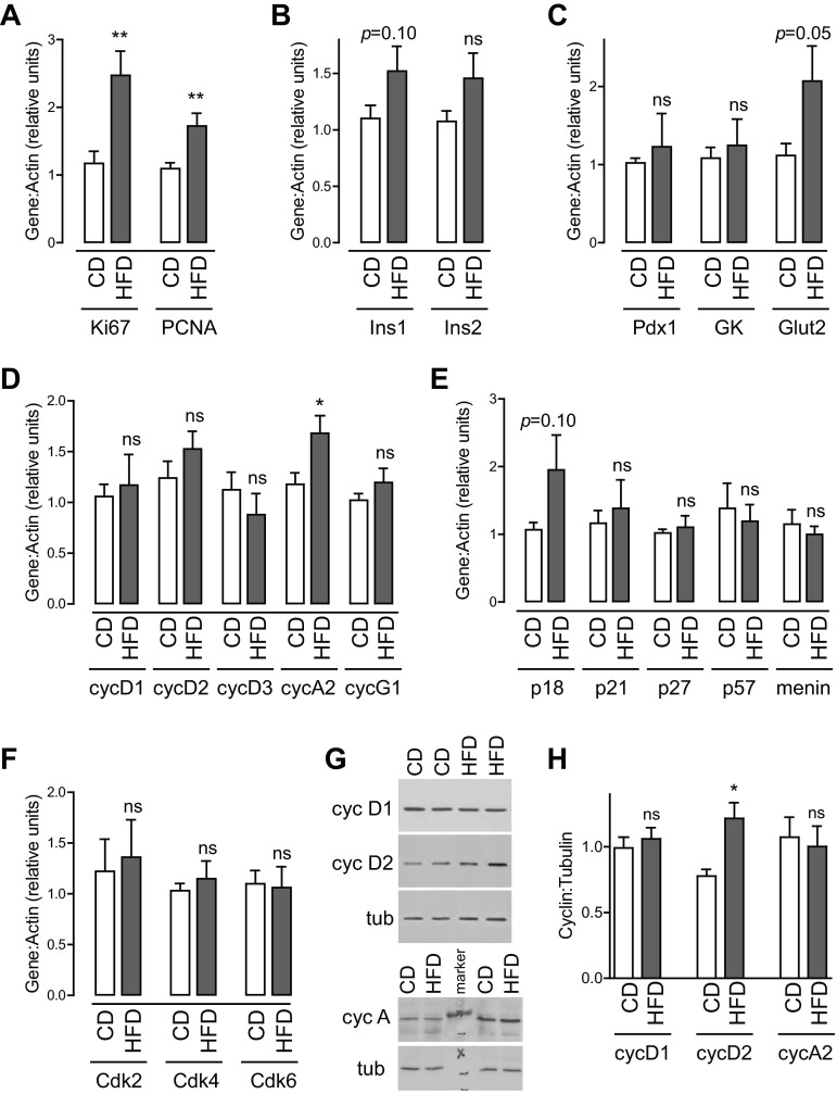
Type 2 diabetes (T2D) is caused by relative insulin deficiency, due in part to reduced β-cell mass (11, 62). Therapies aimed at expanding β-cell mass may be useful to treat T2D (14). Although feeding rodents a high-fat diet (HFD) for an extended period (3-6 mo) increases β-cell mass by inducing β-cell proliferation (16, 20, 53, 54), evidence suggests that adult human β-cells may not meaningfully proliferate in response to obesity. The timing and identity of the earliest initiators of the rodent compensatory growth response, possible therapeutic targets to drive proliferation in refractory human β-cells, are not known. To develop a model to identify early drivers of β-cell proliferation, we studied mice during the first week of HFD exposure, determining the onset of proliferation in the context of diet-related physiological changes. Within the first week of HFD, mice consumed more kilocalories, gained weight and fat mass, and developed hyperglycemia, hyperinsulinemia, and glucose intolerance due to impaired insulin secretion. The β-cell proliferative response also began within the first week of HFD feeding. Intriguingly, β-cell proliferation increased before insulin resistance was detected. Cyclin D2 protein expression was increased in islets by day 7, suggesting it may be an early effector driving compensatory β-cell proliferation in mice. This study defines the time frame and physiology to identify novel upstream regulatory signals driving mouse β-cell mass expansion, in order to explore their efficacy, or reasons for inefficacy, in initiating human β-cell proliferation.

Keywords: diet-induced obesity; islet replication; overnutrition; pancreatic β-cell mitosis; short-term high-fat diet.


Figures

Fig. 1.
Fig. 1.
C57BL/6J mice consumed more kilocalories and gained weight in the first 7 days of high-fat feeding. A and B: mice given high-fat diet (HFD) ate fewer grams of chow per day (A), which nonetheless translated to more kilocalories per day (B) than mice fed control chow (CD) (n = 51–53). C: young adult mice fed CD gained a small amount of body weight over the 7-day intervention, whereas mice fed HFD gained weight as early as day 3, with a 10% weight gain by day 7 (n = 59–60). D: testicular fat pad mass was doubled on day 7 in mice fed HFD (n = 6–7). Data are expressed as means ± SE; P values were calculated by Student's t-test (A, B, D) or ANOVA (C). *P < 0.05, **P < 0.01, ****P < 0.0001; ns, nonsignificant.
Fig. 2.
Fig. 2.
Nonfasting blood glucose and insulin levels increased after 7 days of HFD. Morning tail blood samples were obtained on days 0 and 7 to assess blood glucose, insulin, and free fatty acid (FFA) levels. A: blood glucose increased by day 7 in mice fed HFD, whereas mice fed CD maintained similar glucose levels throughout the experiment (n = 39–40). B: by day 7, HFD mice had nonfasting hyperinsulinemia (n = 22–31). C: plasma FFAs were not altered by either diet (n = 22–32). Data are expressed as means ± SE; P values were calculated by ANOVA with Bonferroni correction for multiple comparisons. *P < 0.05, **P < 0.01, ***P < 0.001.
Fig. 3.
Fig. 3.
Glucose intolerance began within the 1st wk of high-fat feeding without evidence of insulin resistance. A and B: mice given HFD were intolerant to glucose challenge when tested on day 5 (A, n = 4) and day 7 (B, n = 8). C and D: intraperitoneal challenge with 1.5 U/kg insulin revealed no difference in insulin sensitivity between CD and HFD mice when tested on day 5 (C, n = 7–8) or day 7 (D, n = 4). E and F: ITT with a lower dose of insulin (0.75 U/kg) also showed no decrease in insulin sensitivity in mice on HFD at either time point (n = 4). Insets: area under the curve (AUC) for 120-min glucose tolerance test (GTT) or 60-min insulin tolerance test (ITT). Data are expressed as means ± SE; P values by two-way ANOVA with Bonferroni correction for multiple comparisons. *P < 0.05, **P < 0.01, ***P < 0.001, ****P < 0.0001.
Fig. 4.
Fig. 4.
Insulin-secretory response to glucose challenge was mildly impaired after 1 wk of high-fat feeding. A: blood glucose levels measured before and 10 min after intraperitoneal glucose injection were similar between CD and HFD mice on day 8. B: basal plasma insulin levels were higher in HFD mice, but insulin levels after glucose injection were similar between the groups. C and D: when considered as percent increase from baseline, blood glucose increased similarly in CD and HFD mice (C), but plasma insulin increased less in HFD mice (D). E: in vitro glucose-stimulated insulin secretion was impaired in islets isolated after 7 days of HFD exposure. F: islet insulin content was not altered by 7 days of HFD. Data are expressed as means ± SE; P values were calculated by Student's t-test; n = 12 for A–D, n = 4 for EF. *P < 0.05, **P < 0.01, ***P < 0.001.
Fig. 5.
Fig. 5.
β-Cell proliferation increased within the 1st wk of HFD feeding. A: pancreas sections stained for insulin (green), BrdU (red), and DAPI (blue) show that proliferating β-cells were strongly labeled for accurate quantification. These images are from mice treated with BrdU in the drinking water on days 3–7. B: β-cell proliferation was increased on day 4 of HFD as detected by a single intraperitoneal injection of BrdU on the morning of day 4. C: β-cell proliferation was increased in HFD mice when measured after exposure to BrdU continuously on days 3–7. D: pancreas sections stained for insulin (green), PCNA (red), and DAPI (blue) show that proliferating β-cells were strongly labeled for accurate quantification. E: percent insulin-positive cells colabeled for PCNA was increased on day 7; n = 8 (B) and n = 4 (C and E). Data are expressed as means ± SE; P values were calculated by Student's t-test. *P < 0.05, **P < 0.01.
Fig. 6.
Fig. 6.
Compensatory β-cell mass expansion began during the 1st wk of HFD. A–C: β-cell mass had begun to increase after only 7 days of HFD (n = 12; A). This increase was not due to increased pancreas weight (n = 12; B) but instead to an increase in percent pancreatic area made up of islets (n = 12; C). D: size of individual β-cells was not altered by HFD treatment (n = 4). E: analysis of size distribution of pancreatic islets revealed no increase in the number of very small islets (n = 8). F: high-fat feeding increased the proliferation rate to a similar degree in islets of all sizes (n = 4). These data are from mice treated with BrdU in the drinking water on days 3–7. Data are expressed as means ± SE; P values were calculated by Student's t-test (A–D) or two-way ANOVA with Bonferroni correction for multiple comparisons (E: P < 0.0001 for islet size, P = ns for diet; F: P < 0.0001 for diet, P = ns for islet size). **P < 0.01.
Fig. 7.
Fig. 7.
Early compensatory β-cell proliferation is associated with increased cyclin D2 protein expression. Real-time PCR was performed on islets isolated after 7 days of high-fat feeding. A: Ki67 and PCNA expression were increased by HFD, further supporting the conclusion that islet cell proliferation was increased. B and C: analysis of genes related to β-cell function revealed a trend toward an increase in Glut2 expression without alteration of other genes related to β-cell differentiation. D: cyclin A2 mRNA levels were increased after HFD exposure without change in cyclins D1, D2, D3, E1, or G1. E: no changes in G1-S inhibitors were observed between the two diets. F: expression of Cdk genes relevant to the G1-S transition were not altered. G and H: immunoblots showed that cyclin D2 protein was increased in islets after 7 days of HFD; cyclins D1 and A were not; n = 14–19 (A–F); n = 7 (G, H). Data are expressed as means ± SE; P values were calculated by Student's t-test. *P < 0.05, **P < 0.01.

References

    1. Ahren B, Gudbjartsson T, Al-Amin AN, Martensson H, Myrsen-Axcrona U, Karlsson S, Mulder H, Sundler F. Islet perturbations in rats fed a high-fat diet. Pancreas 18: 75–83, 1999 - PubMed
    1. Ahren B, Pacini G. Insufficient islet compensation to insulin resistance vs. reduced glucose effectiveness in glucose-intolerant mice. Am J Physiol Endocrinol Metab 283: E738–E744, 2002 - PubMed
    1. Ahren J, Ahren B, Wierup N. Increased beta-cell volume in mice fed a high-fat diet: a dynamic study over 12 months. Islets 2: 353–356, 2010 - PubMed
    1. Alonso LC, Watanabe Y, Stefanovski D, Lee EJ, Singamsetty S, Romano LC, Zou B, Garcia-Ocana A, Bergman RN, O'Donnell CP. Simultaneous measurement of insulin sensitivity, insulin secretion, and the disposition index in conscious unhandled mice. Obesity (Silver Spring) 20: 1403–1412, 2012 - PMC - PubMed
    1. Alonso LC, Yokoe T, Zhang P, Scott DK, Kim SK, O'Donnell CP, Garcia-Ocana A. Glucose infusion in mice: a new model to induce beta-cell replication. Diabetes 56: 1792–1801, 2007 - PMC - PubMed

Publication types

LinkOut - more resources