. 2013 Jul;30(1):35-42.
doi: 10.3892/or.2013.2473. Epub 2013 May 15.

The benefit of intraperitoneal chemotherapy for the treatment of colorectal carcinomatosis

Affiliations

The benefit of intraperitoneal chemotherapy for the treatment of colorectal carcinomatosis

Valerie Francescutti et al. Oncol Rep. 2013 Jul.

Abstract

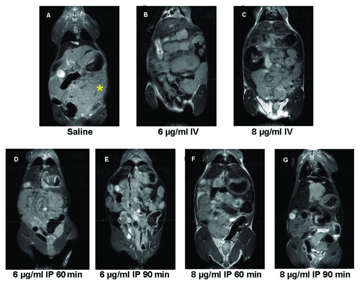
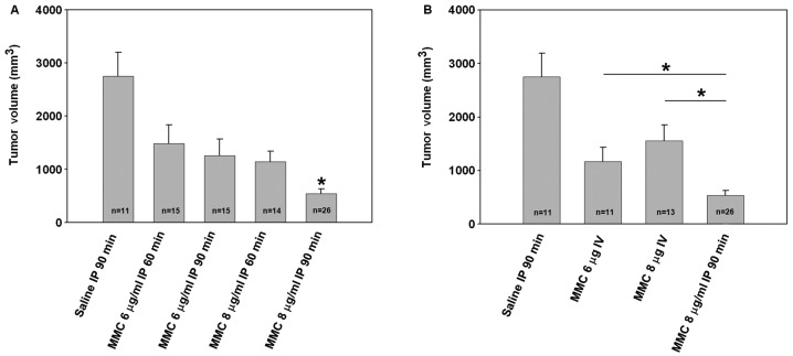
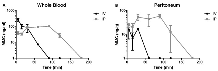
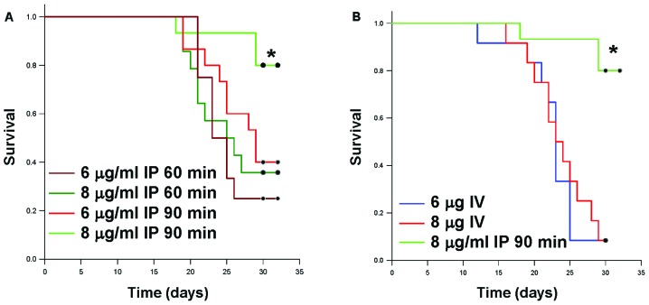
The clinical practice of hyperthermic intraperitoneal chemoperfusion (HIPEC) for carcinomatosis has lacked preclinical justification. A standardized mouse model was created to evaluate the independent effects of intraperitoneal chemotherapy. Diffuse colorectal carcinomatosis was generated in mice prior to intraperitoneal lavage with mitomycin C (MMC) at clinically comparable dosing for variable lengths of time. Tumor volumes, MMC tissue concentrations and survival were measured in comparison to saline lavage and intravenous MMC. Magnetic resonance imaging revealed a direct correlation between tumor volume, MMC dose and exposure time and survival. Intravenous MMC demonstrated a rapid clearance from the blood, lower peritoneal tissue concentrations, less tumor growth inhibition and decreased survival compared to intraperitoneal administration. Intraperitoneal chemotherapy inhibited tumor growth independent of cytoreduction or hyperthermia, demonstrated improved peritoneal tissue concentration and was associated with increased survival. These data support the clinical utility of the intraperitoneal chemotherapy component of HIPEC.


Figures

Figure 1
Figure 1
(A) Coliseum technique with tenting of peritoneum by securing silk sutures to Lego® platform; (B) creation of a petroleum jelly rim to allow complete filling of the abdominal cavity; (C) post-treatment, wicking of chemoperfusate from abdominal cavity using sterile 4 × 4 gauze; (D) abdominal closure with absorbable suture and Vetbond™.
Figure 2
Figure 2
Necropsy images 2, 5, 10, 14 or 21 days after intraperitoneal injection of 5×10⁴ live CT26 cells, showing consistent and progressive (A) small bowel serosal disease and (B) mesenteric disease (magnification, ×8–10) indicated by an asterisk. (C) Frozen section of peritoneum on day 2 after tumor injection demonstrates microscopic tumor implant (asterisk) on the surface of the peritoneum defined by arrows.
Figure 3
Figure 3
The panel of images represent a single slice from coronal T2-weighted MR images for an animal in each experimental group including saline controls. A diffuse pattern of tumor growth in the peritoneum was visualized on the MR images of saline treated control animals with a reduced pattern in the treatment groups. Peritoneal carcinomatosis was well visualized as MRI offered exceptional soft tissue contrast that allowed for accurate, non-invasive volumetric assessments.
Figure 4
Figure 4
(A) Calculated tumor volumes from MR imaging shows a trend toward tumor inhibition associated with increasing intraperitoneal MMC dosing and exposure time compared to saline controls. Mice treated by high dose MMC given for 90 min had a statistically significant decrease in tumor volumes compared to saline controls (P<0.001). (B) Intravenous MMC treated mice trended toward decreased tumor growth compared to saline controls, but a superior antitumor effect is seen when MMC is given intraperitoneally compared to either intravenous treatments (P<0.05).
Figure 5
Figure 5
(A) Whole blood UPLC analysis demonstrates a rapid peak and clearance in MMC concentrations when given intravenously vs. a rapid, but prolonged presence following intraperitoneal delivery. (B) Parietal peritoneum UPLC analysis for MMC shows a similar pattern of rapid peak concentration and quick clearance when given intravenously. Peritoneal MMC concentrations quickly peak and are maintained for a prolonged period even after washout of the peritoneum at 90 min (tissue concentrations presented in logarithmic scale).
Figure 6
Figure 6
Survival is exposure time- and dose-dependent in mice receiving intraperitoneal chemotherapy lavage. (A) Intraperitoneal MMC (8 μg/ml) given for 90 min exhibited a greater improvement in survival compared to both 6 μg/ml for 90 min and 8 μg/ml for 60 min (*P<0.05). (B) Intravenous MMC-treated mice had decreased survival compared to standard HIPEC protocol conditions (i.e. 8 μg/ml intraperitoneal MMC given for 90 min) (*P<0.01).

References

    1. Koppe MJ, Boerman OC, Oyen WJ, Bleichrodt RP. Peritoneal carcinomatosis of colorectal origin: incidence and current treatment strategies. Ann Surg. 2006;243:212–222. - PMC - PubMed
    1. Siegel R, DeSantis C, Virgo K, et al. Cancer treatment and survivorship statistics, 2012. CA Cancer J Clin. 2012;62:220–241. - PubMed
    1. Chu DZ, Lang NP, Thompson C, Osteen PK, Westbrook KC. Peritoneal carcinomatosis in nongynecologic malignancy. A prospective study of prognostic factors. Cancer. 1989;63:364–367. - PubMed
    1. Elias D, Delperro JR, Sideris L, et al. Treatment of peritoneal carcinomatosis from colorectal cancer: impact of complete cytoreductive surgery and difficulties in conducting randomized trials. Ann Surg Oncol. 2004;11:518–521. - PubMed
    1. Sugarbaker PH. A curative approach to peritoneal carcinomatosis from colorectal cancer. Semin Oncol. 2005;32:S68–S73. - PubMed

Publication types