. 2013:4:1849.
doi: 10.1038/ncomms2834.

Coordinated activation of the Rac-GAP β2-chimaerin by an atypical proline-rich domain and diacylglycerol

Affiliations

Coordinated activation of the Rac-GAP β2-chimaerin by an atypical proline-rich domain and diacylglycerol

Alvaro Gutierrez-Uzquiza et al. Nat Commun. 2013.

Abstract

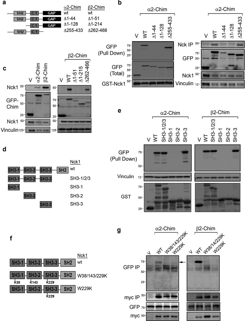
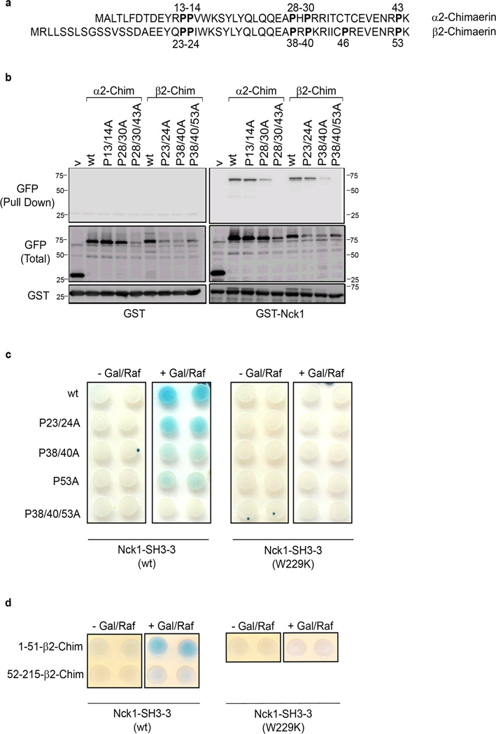
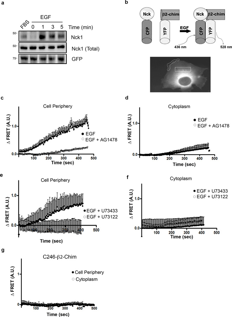
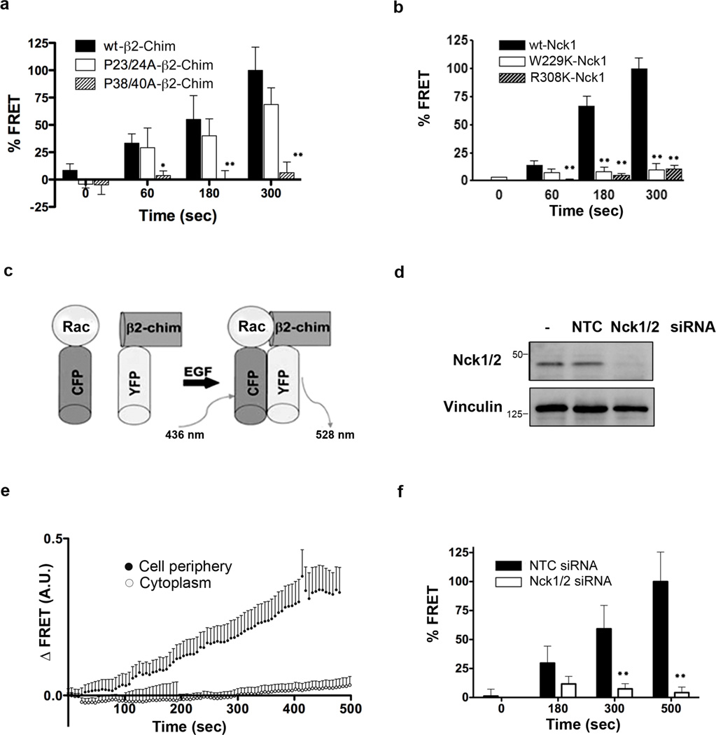
Chimaerins, a family of GTPase activating proteins for the small G-protein Rac, have been implicated in development, neuritogenesis and cancer. These Rac-GTPase activating proteins are regulated by the lipid second messenger diacylglycerol generated by tyrosine kinases such as the epidermal growth factor receptor. Here we identify an atypical proline-rich motif in chimaerins that binds to the adaptor protein Nck1. Unlike most Nck1 partners, chimaerins bind to the third SH3 domain of Nck1. This association is mediated by electrostatic interactions of basic residues within the Pro-rich motif with acidic clusters in the SH3 domain. Epidermal growth factor promotes the binding of β2-chimaerin to Nck1 in the cell periphery in a diacylglycerol-dependent manner. Moreover, β2-chimaerin translocation to the plasma membrane and its peripheral association with Rac1 requires Nck1. Our studies underscore a coordinated mechanism for β2-chimaerin activation that involves lipid interactions via the C1 domain and protein-protein interactions via the N-terminal proline-rich region.


Figures

Figure 1
Figure 1. Interaction of the α2- and β2-chimaerin N-terminal domain with the Nck1 SH3-3 domain
(a) Schematic representation of α2- and β2-chimaerin mutants. (b) Left panel, extracts from COS-1 expressing GFP-α2-chimaerin (wt or mutants) were incubated with GST-Nck1 and pulled down with glutathione Sepharose 4B beads. Levels of chimaerins in the GST precipitates were determined with an anti-GFP antibody. Right panel, extracts of COS-1 cells expressing α2-chimaerin (wt or mutant) were subject to IP with an anti-GFP antibody. Endogenous Nck1 was detected in the IP by Western blot. (c) Left panel, preferential association of β2-chimaerin with endogenous Nck1. Right panel, extracts of COS-1 cells expressing β2-chimaerin (wt or mutant) were subject to IP with an anti-GFP antibody. Endogenous Nck1 was detected in the IP by Western blot. (d) Schematic representation of Nck1 deleted mutants. (e) COS-1 cells expressing GFP-α2-chimaerin (left panel) or GFP-β2-chimaerin (right panel) were incubated with GST, GST-Nck1 or the indicated GST-Nck1 mutants. After pull-down, levels of chimaerins in the GST precipitates were determined with an anti-GFP antibody. (f) Schematic representation of Nck1 SH3 domain point mutants. (g) COS-1 cells were co-transfected with pEGFP-α2-chimaerin (left panel) or pEGFP-β2-chimaerin (right panel) together with plasmids encoding myc-tagged Nck1 (wt or mutants). Twenty four hr later cells were subject to IP with an anti-myc tag antibody. α2- and β2-chimaerins in the IP (arrow) were detected by Western blot using an anti-GFP antibody. In all panels, similar results were observed in at least 3 independent experiments.
Figure 2
Figure 2. Identification of an atypical Pro-rich region in α2- and β2-chimaerin
(a) Alignment of α2- and β2-chimaerin N-terminal regions. (b) Extracts from COS-1 cells expressing GFP-α2- or GFP-β2-chimaerin (wt or Pro→Ala mutants) were incubated with GST (left panel) or GST-Nck1 (right panel). Chimaerins in GST precipitates were detected by Western blot with an anti-GFP antibody. (c) Association by yeast-two hybrid between wild-type or mutated β2-chimaerin (amino acids 1–186) and SH3-3-Nck1 Wt or W229K mutant. Gal/Raf, galactosidase/raffinose. (d) Interaction between the N-terminal region of β2-chimaerin (amino acids 1–53) and SH3-3-Nck1. In all panels, similar results were observed in at least 3 independent experiments.
Figure 3
Figure 3. Basic amino acids in the Pro-rich region of β2-chimaerin mediate the interaction with Nck1 SH3-3
(a) Analysis of binding interfaces between the third Nck1 domain (SH3-3) and the Pro-rich motif PRPKR (β2-chimaerin). White = neutral charge; red = negative charge; blue = positive charge; yellow = PRPKR (β2-chimaerin); green = PHPRR (α2-chimaerin). (b) COS-1 cells were transfected with pEGFP plasmids encoding wt-β2-chimaerin or various mutated forms of β2-chimaerin. After 24 h cell extracts were subject to IP with an anti-GFP antibody. Nck1 in the IP were detected by Western blot. Two additional experiments gave similar results. (c) Representative Biacore sensorgrams showing the interaction between immobilized GST-Nck1 SH3-3 or GST alone and increasing concentrations (1, 10, 20, 40 µM) of wild-type β2-chimaerin N-terminal peptides. (d) Representative Biacore sensorgrams showing the interaction between immobilized GST-Nck1 SH3-3 and increasing concentrations (1, 10, 20, 40 µM) of mutated β2-chimaerin N-terminal peptides.
Figure 4
Figure 4. The interaction between β2-chimaerin and Nck1 depends on the membrane-bound conformation of β2-chimaerin
(a) Docking model of β2-chimaerin and Nck1 SH3-3 to show that steric clash makes binding of Nck-SH3 to the closed state of β2-chimaerin unfavorable. β2-chimaerin is shown as ribbon and translucent surface. Nck1 SH3-3 is shown as purple ribbon. (b) Enhanced view of (a). (c) β2-chimaerin binding to PMA-containing SLVs. S = supernatant fraction, P = pellet fraction. (d) Binding curve for PMA-dependent recruitment of β2-chimaerin. (e) Nck1 SH3-3 binding to PMA-containing SLVs (f) Co-recruitment of Nck1 SH3-3 and β2-chimaerin to PMA-containing SLVs. (g) Binding curve for Nck1 SH3-3 interaction with β2-chimaerin gives a KD = 1.7 µM. Results are the mean ± S.D. of 3 experiments.
Figure 5
Figure 5. EGF promotes the interaction between β2-chimaerin and Nck1 in a DAG-dependent manner
(a) COS-1 cells expressing GFP-β2-chimaerin were serum-starved for 24 hr and treated with EGF (100 ng/ml). Cell extracts were subject to IP with an anti-GFP antibody. Nck1 was detected in the IP using an anti-Nck antibody. Lane 1, cells growing in medium with 10% FBS. (b) Schematic representation of the FRET assay to assess Nck1-β2-chimaerin interaction in cells. Typical peripheral and cytoplasmatic cell sections selected for analysis are shown. (c) FRET in peripheral cellular sections was measured every 3 sec after EGF treatment (100 ng/ml), both in the absence and presence of the EGFR inhibitor AG1478 (1 µM). (d) FRET in cytoplasmatic cellular sections after EGF treatment, both in the absence and presence of AG1478. (e) and (f) FRET in cell peripheral (e) and cytoplasmatic (f) sections after EGF treatment in the presence of either U73122 or its inactive analog U73433 (10 µM, added 30 min before and during EGF treatment). (g) FRET in peripheral and cytoplasmatic sections using the pair CFP-Nck1 and YFP-C246A-β2-chimaerin. For all FRET experiments, data are expressed as mean ± S.E.M. of 6–13 cells.
Figure 6
Figure 6. Nck is required for β2-chimaerin activation by EGF
(a) FRET between YFP-β2-chimaerin (wt, and mutants P23/24A and P38/40A) and CFP-Nck1 after treatment of COS-7 cells with EGF (100 ng/ml). ). Data (mean ± S.E.M.) are expressed as % of maximum FRET with wt YFP-β2-chimaerin (ANOVA test with Bonferroni adjustment was used; *p<0.05; **p<0.01; n=10). (b) FRET between YFP-β2-chimaerin and CFP-Nck1 (wt, W229K or W308K) after treatment of COS-7 cells with EGF (100 ng/ml). Data (mean ± S.E.M.) are expressed as % of maximum FRET with wt CFP-Nck1 (ANOVA test with Bonferroni adjustment was used; *p<0.05; **p<0.01; n=10). (c) Schematic representation of the FRET assay to assess Rac1-β2-chimaerin association (adapted from ). (d) Depletion of Nck1/2 in COS-7 cells using RNAi, as determined 48 h after transfection. NTC, non-target control siRNA. (e) FRET analysis in COS-7 cells expressing CFP-Rac1 and YFP-β2-chimaerin was measured every 6 s after EGF treatment, both in peripheral and cytoplasmatic sections. Data are expressed as mean ± S.E.M. (n= 10). (f) FRET in response to EGF in the periphery of cells transfected with either NTC or Nck1/2 RNAi. Data (mean ± S.E.M.) are expressed as percentage of maximum FRET with wt YFP-β2-chimaerin (unpaired t-student test was used; *p<0.05; **p<0.01; n=10).
Figure 7
Figure 7. Nck is required for β2-chimaerin activation by sodium pervanadate
(a) COS-7 cells were transfected with pEGFP-β2-chimaerin and either Nck1/2 or non-target control (NTC) siRNA. After 48 hr cells were treated with PMA (10 nM, 15 min) either in the absence or presence of sodium pervanadate (1 mM, 15 min before and during PMA), and visualized by confocal microscopy. Representative micrographs are shown. (b) Quantification of translocation using ImageJ. Data are expressed as mean ± S.E.M. of 7–15 individual cells for each group. (unpaired t-student test was used; **p<0.01 vs. NTC siRNA). NTC, non-target control. (c) FRET analysis in COS-7 cells expressing CFP-Nck1 and YFP-β2-chimaerin was measured after sodium pervanadate treatment, both in peripheral and cytoplasmatic sections. Data are expressed as mean ± S.E.M. (n= 10). (d) FRET between YFP-β2-chimaerin and Nck1 (wt, W229K or W308K) at different times after treatment with sodium pervanadate (1 mM). Data (mean ± S.E.M.) are expressed as percentage of maximum FRET with wt YFP-β2-chimaerin. (ANOVA test with Bonferroni adjustment was used;*p<0.05; **p<0.01; n=10) (e) Model for EGFR-mediated regulation of β2-chimaerin via DAG and Nck1. Size bar: 10 µm.

References

    1. Caloca MJ, Delgado P, Alarcon B, Bustelo XR. Role of chimaerins, a group of Rac-specific GTPase activating proteins, in T-cell receptor signaling. Cell Signal. 2008;20:758–770. - PMC - PubMed
    1. Wegmeyer H, Egea J, Rabe N, Gezelius H, Filosa A, Enjin A, et al. EphA4-dependent axon guidance is mediated by the RacGAP alpha2-chimaerin. Neuron. 2007;55:756–767. - PubMed
    1. Beg AA, Sommer JE, Martin JH, Scheiffele P. alpha2-Chimaerin is an essential EphA4 effector in the assembly of neuronal locomotor circuits. Neuron. 2007;55:768–778. - PubMed
    1. Suliman SG, Stanik J, McCulloch LJ, Wilson N, Edghill EL, Misovicova N, et al. Severe insulin resistance and intrauterine growth deficiency associated with haploinsufficiency for INSR and CHN2: new insights into synergistic pathways involved in growth and metabolism. Diabetes. 2009;58:2954–2961. - PMC - PubMed
    1. Colon-Gonzalez F, Leskow FC, Kazanietz MG. Identification of an autoinhibitory mechanism that restricts C1 domain-mediated activation of the Rac-GAP alpha2-chimaerin. J Biol Chem. 2008;283:35247–35257. - PMC - PubMed

Publication types

MeSH terms