

Burn injury enhances bone formation in heterotopic ossification model
- PMID: 23673767
- PMCID: PMC4498401
- DOI: 10.1097/SLA.0b013e318291da85
Burn injury enhances bone formation in heterotopic ossification model
Abstract
Objective: To demonstrate the pro-osteogenic effect of burn injury on heterotopic bone formation using a novel burn ossicle in vivo model.
Background: Heterotopic ossification (HO), or the abnormal formation of bone in soft tissue, is a troubling sequela of burn and trauma injuries. The exact mechanism by which burn injury influences bone formation is unknown. The aim of this study was to develop a mouse model to study the effect of burn injury on heterotopic bone formation. We hypothesized that burn injury would enhance early vascularization and subsequent bone formation of subcutaneously implanted mesenchymal stem cells.
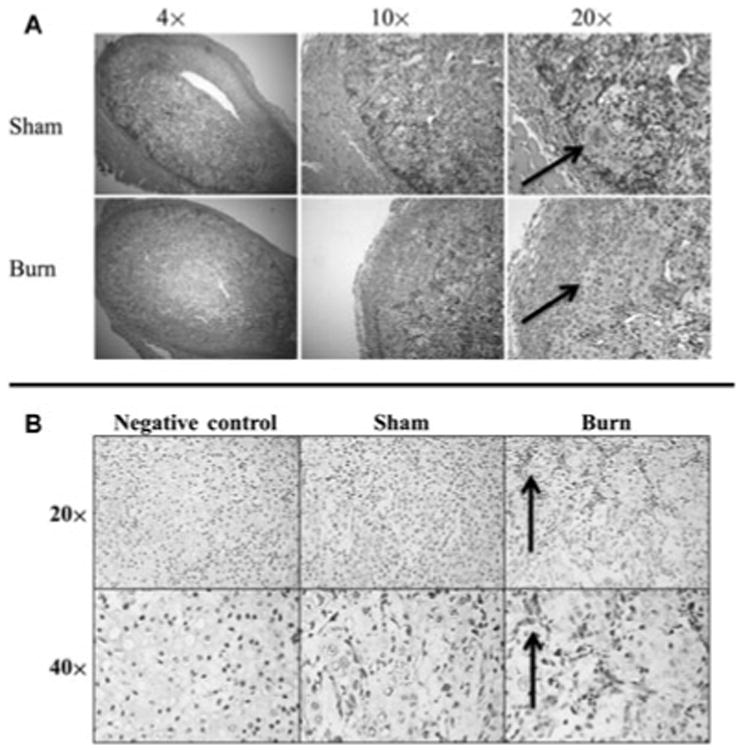
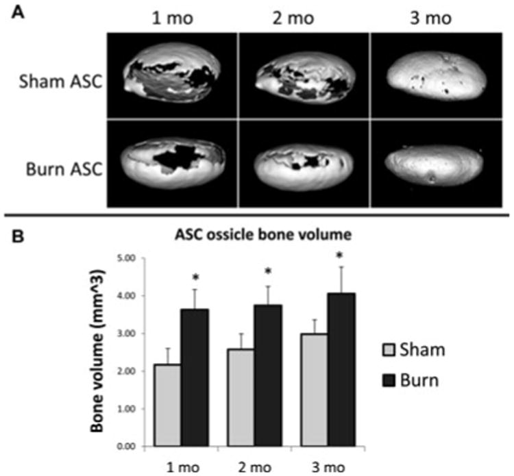
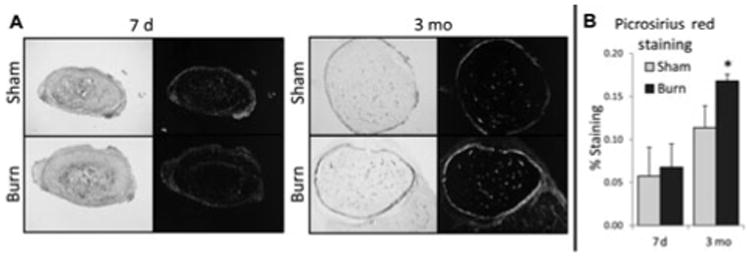
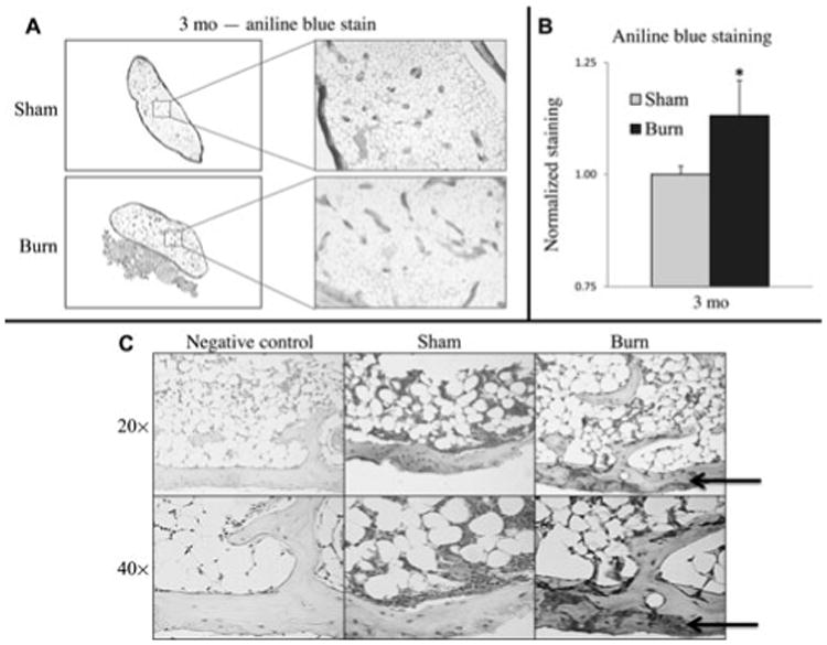
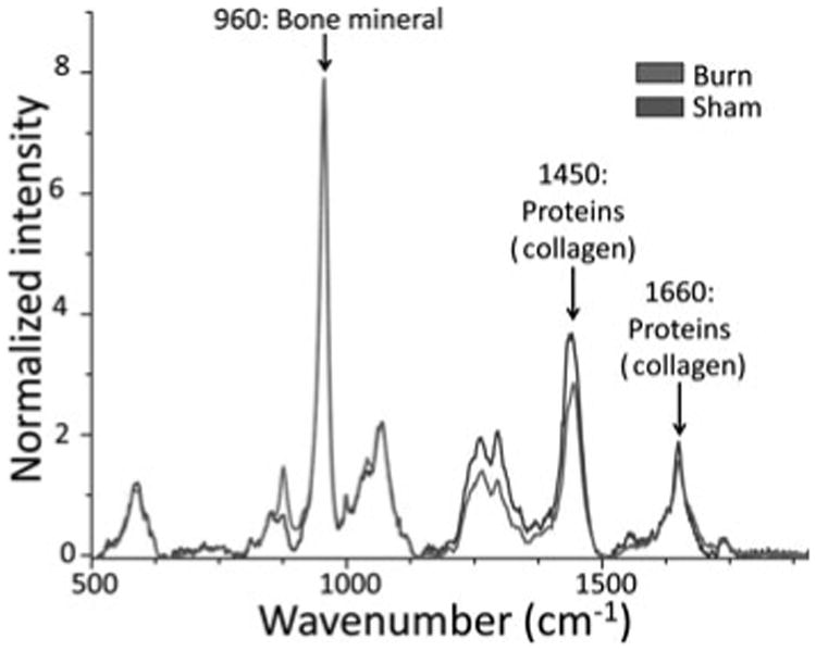
Methods: Mouse adipose-derived stem cells were harvested from C57/BL6 mice, transfected with a BMP-2 adenovirus, seeded on collagen scaffolds (ossicles), and implanted subcutaneously in the flank region of 8 adult mice. Burn and sham groups were created with exposure of 30% surface area on the dorsum to 60°C water or 30°C water for 18 seconds, respectively (n = 4/group). Heterotopic bone volume was analyzed in vivo by micro-computed tomography for 3 months. Histological analysis of vasculogenesis was performed with platelet endothelial cell adhesion molecule staining. Osteogenic histological analysis was performed by Safranin O, Picrosirius red, and aniline blue staining. Qualitative analysis of heterotopic bone composition was completed with ex vivo Raman spectroscopy.
Results: Subcutaneously implanted ossicles formed heterotopic bone. Ossicles from mice with burn injuries developed significantly more bone than sham control mice, analyzed by micro-computed tomography at 1, 2, and 3 months (P < 0.05), and had enhanced early and late endochondral ossification as demonstrated by Safranin O, Picrosirius red, and aniline blue staining. In addition, burn injury enhanced vascularization of the ossicles (P < 0.05). All ossicles demonstrated chemical composition characteristic of bone as demonstrated by Raman spectroscopy.
Conclusions: Burn injury increases the predilection to osteogenic differentiation of ectopically implanted ossicles. Early differences in vascularity correlated with later bone development. Understanding the role of burn injury on heterotopic bone formation is an important first step toward the development of treatment strategies aimed to prevent unwanted and detrimental heterotopic bone formation.
Figures

References
-
- Chen HC, Yang JY, Chuang SS, et al. Heterotopic ossification in burns: our experience and literature reviews. Burns. 2009;35:857–862. - PubMed
-
- Shimono K, Morrison TN, Tung WE, et al. Inhibition of ectopic bone formation by a selective retinoic acid receptor alpha-agonist: a new therapy for heterotopic ossification? J Orthop Res. 2010;28:271–277. - PubMed
-
- Potter BK, Forsberg JA, Davis TA, et al. Heterotopic ossification following combat-related trauma. J Bone Joint Surg Am. 2010;92(suppl 2):74–89. - PubMed
-
- Ji Y, Christopherson GT, Kluk MW, et al. Heterotopic ossification following musculoskeletal trauma: modeling stem and progenitor cells in their microenvironment. Adv Exp Med Biol. 2011;720:39–50. - PubMed
-
- Tsionos I, Leclercq C, Rochet JM. Heterotopic ossification of the elbow in patients with burns. Results after early excision. J Bone Joint Surg Br. 2004;86:396–403. - PubMed
MeSH terms
Grants and funding
LinkOut - more resources
Full Text Sources
Other Literature Sources
Medical
Molecular Biology Databases
