. 2013 Sep;38(10):1889-98.
doi: 10.1038/npp.2013.122. Epub 2013 May 15.

Hippocampal network connectivity and activation differentiates post-traumatic stress disorder from generalized anxiety disorder

Affiliations

Hippocampal network connectivity and activation differentiates post-traumatic stress disorder from generalized anxiety disorder

Ashley C Chen et al. Neuropsychopharmacology. 2013 Sep.

Abstract

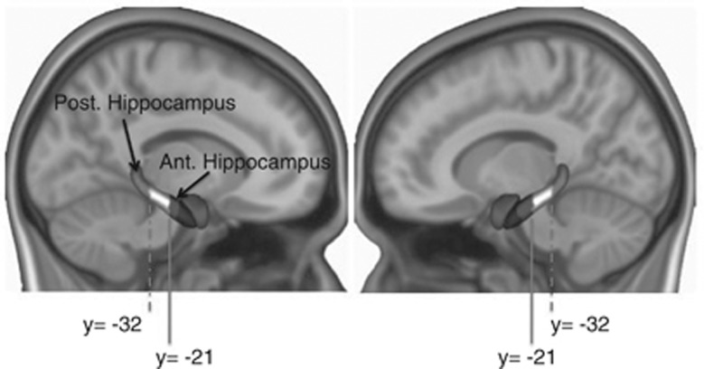
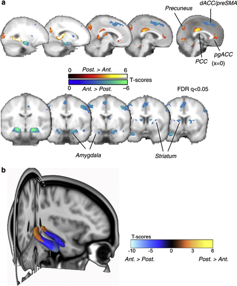
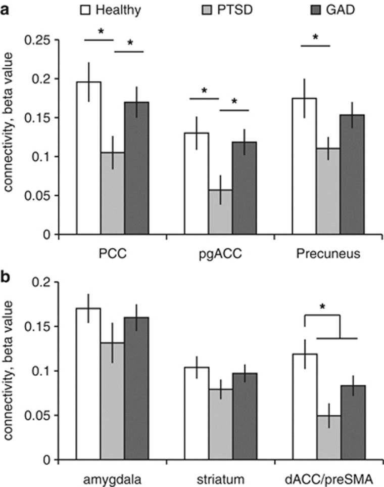
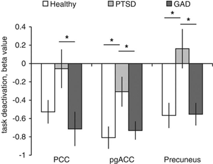
Anxiety disorders are a diverse group of clinical states. Post-traumatic stress disorder (PTSD) and generalized anxiety disorder (GAD), eg, share elevated anxiety symptoms, but differ with respect to fear-related memory dysregulation. As the hippocampus is implicated in both general anxiety and fear memory, it may be an important brain locus for mapping the similarities and differences among anxiety disorders. Anxiety and fear also functionally associate with different subdivisions of the hippocampus along its longitudinal axis: the human posterior (rodent dorsal) hippocampus is involved in memory, through connectivity with the medial prefrontal-medial parietal default-mode network, whereas the anterior (rodent ventral) hippocampus is involved in anxiety, through connectivity with limbic-prefrontal circuits. We examined whether differential hippocampal network functioning may help account for similarities and differences in symptoms in PTSD and GAD. Network-sensitive functional magnetic resonance imaging-based resting-state intrinsic connectivity methods, along with task-based assessment of posterior hippocampal/default-mode network function, were used. As predicted, in healthy subjects resting-state connectivity dissociated between posterior hippocampal connectivity with the default-mode network, and anterior hippocampal connectivity to limbic-prefrontal circuitry. The posterior hippocampus and the associated default-mode network, across both resting-state connectivity and task-based measures, were perturbed in PTSD relative to each of the other groups. By contrast, we found only modest support for similarly blunted anterior hippocampal connectivity across both patient groups. These findings provide new insights into the neural circuit-level dysfunctions that account for similar vs different features of two major anxiety disorders, through a translational framework built on animal work and carefully selected clinical disorders.


Figures

Figure 1
Figure 1
Illustration of the seed regions of interest (ROI). Bilateral anterior and posterior hippocampal subdivisions (see arrows) were used in the functional connectivity analyses. Notably, we excluded voxels with any probability of being in the amygdala (black) from the anterior hippocampus ROI.
Figure 2
Figure 2
Dissociable neural circuits within the human hippocampus revealed by resting-state functional connectivity. (a) Distinct connectivity profiles of the anterior and posterior hippocampus in humans mirrored anatomical connectivity of the ventral and dorsal hippocampus in experimental animals. Although the anterior hippocampus was preferentially connected to subcortical limbic structures and dorsal anterior cingulate cortex (dACC)/pre-supplementary motor area (pre-SMA) (blue), the posterior hippocampus was more connected to the memory-related default-mode network (red). Displays are in neurological convention. MNI x-coordinates of the sagital slices on the top row are: −26, −13, −6, −3, and 0, respectively. MNI y-coordinates of the coronal slices at the bottom row are: −14, −4, −2, 6, and 8, respectively. (b) Findings from a connectivity analysis, seeding with the ‘targets' of each subdivision (defined by a separate cohort), revealed a parcellation, along the anterior–posterior axis, within the hippocampus. PCC, posterior cingulate cortex; pgACC, pregenual anterior cingulate cortex; preCG, precentral gyrus.
Figure 3
Figure 3
Posterior and anterior hippocampal resting-state functional connectivity in matched cohorts of healthy individuals and patients with post-traumatic stress disorder (PTSD) and generalized anxiety disorder (GAD). (a) Group comparisons for the posterior hippocampus show that diminished posterior hippocampal connectivity is specific to PTSD (asterisk ‘*' signifies a group difference at p<0.05). (b) Group comparisons for the anterior hippocampus suggest a general impairment in anxiety disorders (asterisk ‘*' signifies a group difference (healthy vs PTSD or GAD) at p<0.05). Plotted are mean beta estimates±SE. dACC, dorsal anterior cingulate cortex; PCC, posterior cingulate cortex; pgACC, pregenual anterior cingulate cortex; pre-SMA, pre-supplementary motor area.
Figure 4
Figure 4
Task-independent deactivation in the posterior hippocampus/default-mode network during attention-demanding tasks. The degree of task-independent deactivation in the same default-mode network components that in the resting state were preferentially connected with the posterior hippocampus was blunted only in post-traumatic stress disorder (PTSD) patients, relative to healthy individuals, as well as generalized anxiety disorder (GAD) patients. Asterisk ‘*' signifies a group difference at p<0.05. Plotted are mean beta estimates±SE. PCC, posterior cingulate cortex; pgACC, pregenual anterior cingulate cortex.

References

    1. Adhikari A, Topiwala MA, Gordon JA. Synchronized activity between the ventral hippocampus and the medial prefrontal cortex during anxiety. Neuron. 2010;65:257–269. - PMC - PubMed
    1. Aggleton JP, Wright NF, Vann SD, Saunders RC. Medial temporal lobe projections to the retrosplenial cortex of the macaque monkey. Hippocampus. 2012;22:1883–1900. - PMC - PubMed
    1. Amaral DG. Handbook of Physiology, the Nervous System. Waverly Press: Baltimore; 1987. Memory: anatomical organization of candidate brain regions; pp. 211–294.
    1. American Psychiatric Association 1994Diagnostic and Statistical Manual of Mental Disorders4th edn.American Psychiatric Association: Washington, DC
    1. Amunts K, Kedo O, Kindler M, Pieperhoff P, Mohlberg H, Shah NJ, et al. Cytoarchitectonic mapping of the human amygdala, hippocampal region and entorhinal cortex: intersubject variability and probability maps. Anat Embryol (Berl) 2005;210:343–352. - PubMed

Publication types