. 2013 Sep;74(3):458-71.
doi: 10.1002/ana.23815. Epub 2013 May 14.

Adoptive regulatory T-cell therapy protects against cerebral ischemia

Affiliations

Adoptive regulatory T-cell therapy protects against cerebral ischemia

Peiying Li et al. Ann Neurol. 2013 Sep.

Abstract

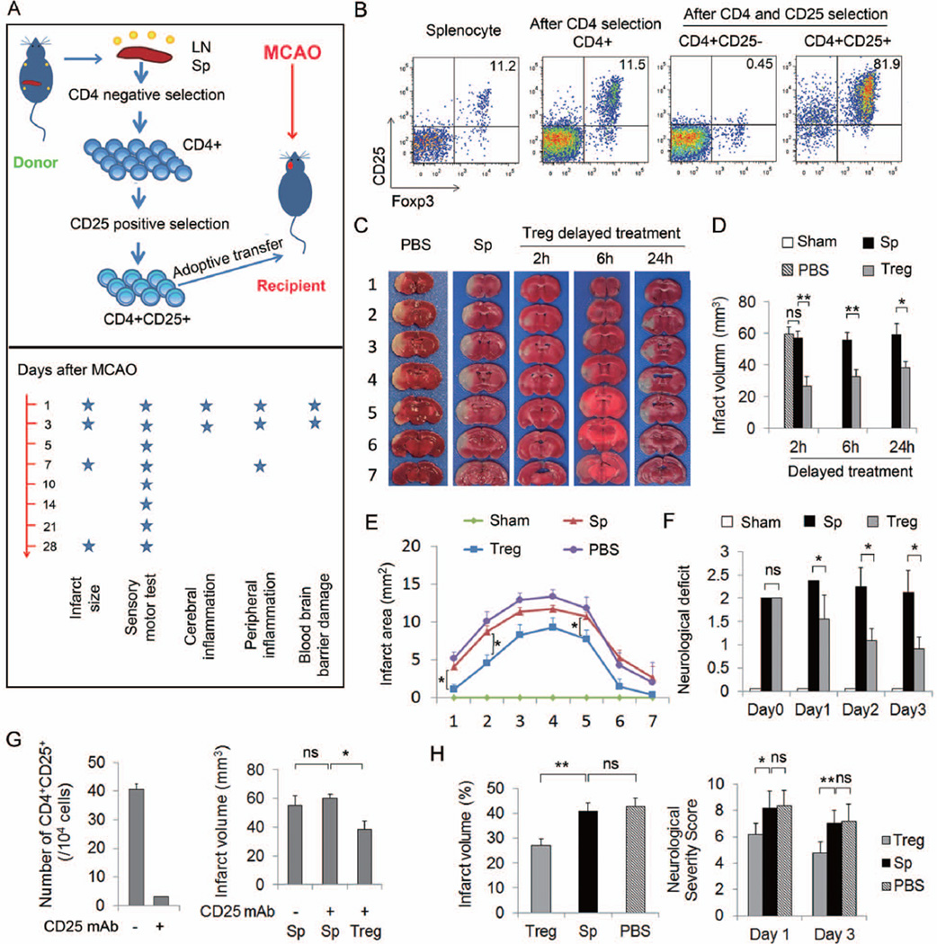
Objective: Recent evidence suggests that functional deficiency in regulatory T cells (Tregs), an innate immunomodulator, exacerbates brain damage after cerebral ischemia. We therefore evaluated the effect of Treg transfer in rodent models of ischemic stroke and further investigated the mechanism underlying Treg-afforded neuroprotection.

Methods: We examined the therapeutic potential of Tregs and the mechanisms of neuroprotection in vivo in 2 rodent models of ischemic stroke and in vitro in Treg-neutrophil cocultures using a combined approach including cell-specific depletion, gene knockout mice, and bone marrow chimeras.

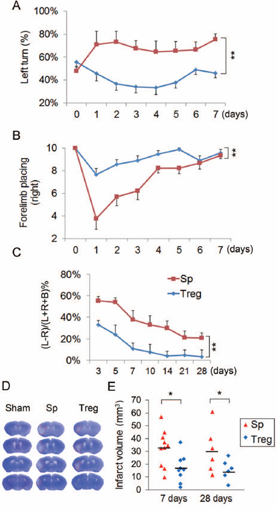
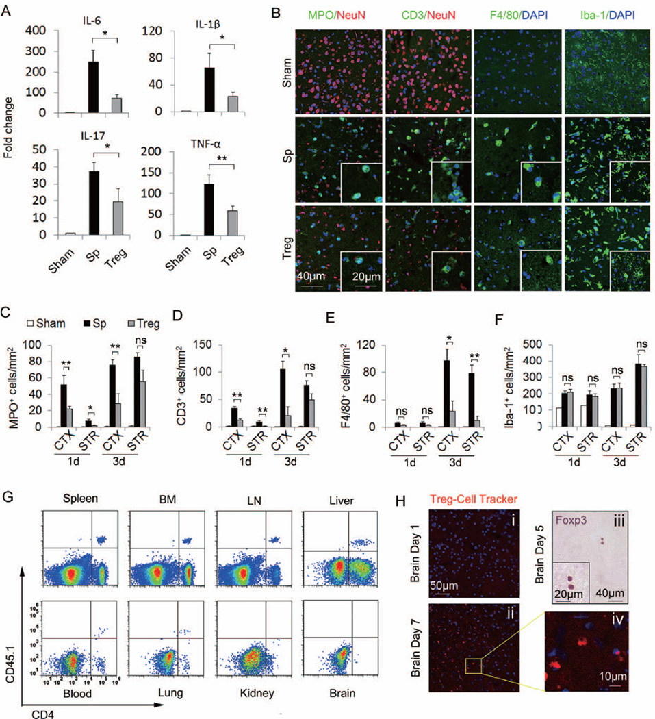
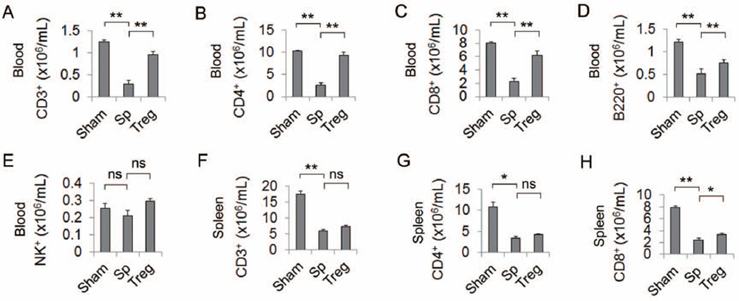
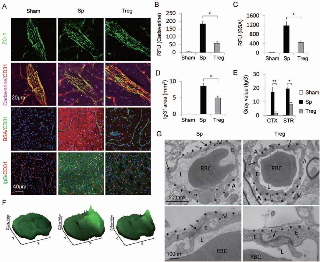
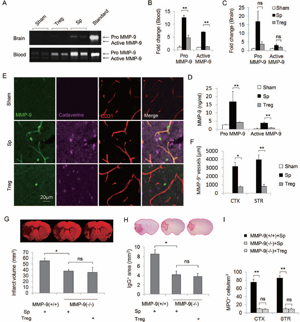
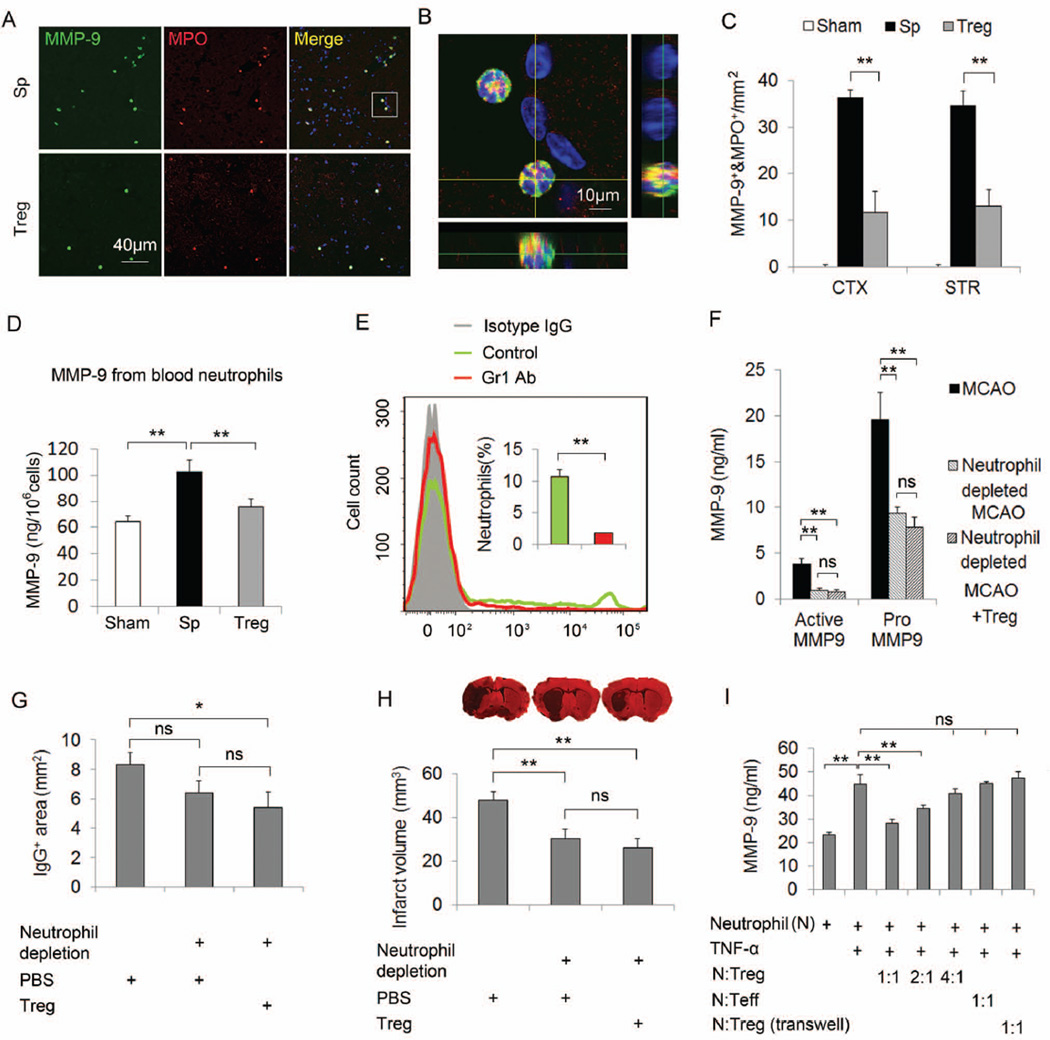
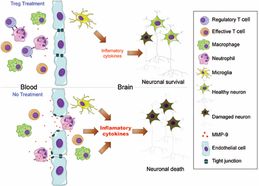
Results: Systemic administration of purified Tregs at 2, 6, or even 24 hours after middle cerebral artery occlusion resulted in a marked reduction of brain infarct and prolonged improvement of neurological functions lasting out to 4 weeks. Treg-afforded neuroprotection was accompanied by attenuated blood-brain barrier (BBB) disruption during early stages of ischemia, decreased cerebral inflammation, and reduced infiltration of peripheral inflammatory cells into the lesioned brain. Surprisingly, Tregs exerted early neuroprotection without penetrating into the brain parenchyma or inhibiting the activation of residential microglia. Rather, both in vivo and in vitro studies demonstrated that Tregs suppressed peripheral neutrophil-derived matrix metallopeptidase-9 production, thus preventing proteolytic damage of the BBB. In addition to its potent central neuroprotection, Treg treatment was shown to ameliorate poststroke lymphopenia, suggesting a beneficial effect on immune status.

Interpretation: Our study suggests that Treg adoptive therapy is a novel and potent cell-based therapy targeting poststroke inflammatory dysregulation and neurovascular disruption.


Figures

Figure 1
Figure 1. Adoptive transfer of Tregs confers neuroprotection against focal cerebral ischemia
(A) Scheme for experimental design. Tregs or splenocytes were isolated from pooled spleens and lymph nodes of donors and injected intravenously (2×10⁶ cells/animal) into recipients at 2, 6 or 24 h after MCAO. Time line for parameter measurements is indicated. (B) Representative flow cytometry plots of CD25 and Foxp3 expression on splenocyte, CD4+ T cells after negative selection, CD4+CD25 and CD4+CD25+ subsets of T cells after double selection. (C–E) Treg-afforded protection in mice at 3d after 60 min MCAO. (C) Representative TTC stained coronal sections showed a smaller cerebral infarct in a mouse with adoptively-transferred Tregs than in a splenocyte-transferred or PBS-treated mouse. (D) Infarct volumes in mice treated with Tregs at 2h (n=7/group), 6h (n=8–9/group) or 24h (n=6–7/group) after MCAO were significantly reduced. (E) Infarct areas of 7 consecutive coronal sections, 1 mm apart, throughout the MCA territory in mice that received treatments at 2h after MCAO (n=7/group). (F) Treg treatment with 2h delay improved neurological deficits in mice over 3d after MCAO compared to splenocyte- or PBS-treatment (n=7/group). (G) Exogenous Tregs protected the ischemic brain in the absence of endogenous Tregs. Mice were injected intraperitoneally (i.p). with either PBS (control) or 300 µg of CD25-specific antibody (CD25 mAb) 2d prior to MCAO. Left: Flow cytometry analysis showing that endogenous Tregs were depleted in anti-CD25 mAb treated mice. Right: Infarct volume was measured 3d after MCAO. (H) Treg-afforded protection in rats at 3d after MCAO. Left: Infarct volume in rats treated with Tregs, splenocytes or PBS at 2h after 120 min of MCAO. Right: Treg-treated rats demonstrated reduced neurological severity scores at 1d and 3d after MCAO compared to splenocyte- or PBS-treated rats (n=5/group). Data are mean ± SE. *P<0.05, **P<0.01.
Figure 2
Figure 2. Tregs confer long-term neuroprotection against cerebral ischemia
Tregs or splenocytes were transferred intravenously into recipient mice at 2h after MCAO. Brain infarct and sensorimotor functions were assessed up to 28d after MCAO. (A–B) Acute sensorimotor dysfunction at 1–7d after ischemia was significantly improved in Treg-treated mice as assessed by the corner test (A, n=10/group) and forelimb placing test (B, n=10/group). Corner test performance was expressed by the percentage of left turns out of 10 turn trials. The performance in forelimb placing test was expressed as the number of successful placing responses on the impaired right forelimb out of 10 placing trials. (C) Treg-treated mice demonstrated improved long-term sensorimotor performance as assessed by cylinder test up to 28d after ischemia (n=6/group). The number of left, right, or both forepaw contacts were counted, and the performance asymmetry was expressed as (left−right)/(left+right+both)×100% paw use in 5 min. (D) Representative cresyl violet stained brain sections obtained at 7d after MCAO showed smaller infarct in a Treg-treated mouse than in a splenocyte-transferred mouse. (E) Infarct volume determined at 7d (n=10/group) and 28d (n=6/group) after ischemia with cresyl violet staining and MAP2 staining, respectively, was significantly reduced in Treg-treated mice. Data are mean ± SE. *P<0.05, **P<0.01.
Figure 3
Figure 3. Tregs attenuate post-ischemic inflammation and reduce the early infiltration of peripheral immune cells into the brain ahead of their own CNS infiltration
(A) Quantitative real-time PCR for mRNA expression of IL-6, IL-1β, IL-17, and TNF-α in the ischemic hemispheres from animals with 60 min MCAO and 24h reperfusion (n=6/group). IL-6, IL-1β, IL-17, and TNF-α mRNA were significantly decreased in Treg-treated mice compared to splenocyte-treated mice. (B) Representative immunofluorescent staining of MPO, CD3, F4/80, and Iba-1 on brain sections obtained 3d after MCAO. (C-F) Time courses for the infiltration of MPO+ neutrophilic granulocytes (C), CD3+ T cells (D), F4/80+ macrophages (E) and the activation of Iba-1+ microglial cells (F) in the ischemic brains of Treg-treated mice compared to splenocyte-treated and sham-operated mice (n=6/group). (G) Adoptively transferred CD45.1+CD4+ Tregs present in the spleen, bone marrow (BM), lymph node (LN), liver, blood and lung, but not in the brain or kidney at 1d after MCAO. Plots are representative of four animals. (H) Delayed brain infiltration of Tregs after stroke. Cell tracker-labeled Tregs were observed in the brain at 7d (ii, iv) but not 1d (i) after MCAO. (iii) Immunohistochemical staining of Foxp3 in brain sections at 5d after MCAO. Images are representative of sections from four animals. Data are mean ± SE. *P<0.05, **P<0.01.
Figure 4
Figure 4. Tregs did not exacerbate post-stroke immunosuppression
(A–E) Treg treatment did not exacerbate the immunosuppression in the blood after MCAO. At 5d after stroke, blood cells were isolated and stained for CD3+ T cells (A), CD4+ T helper cells (B), CD8+ T cytotoxic cells (C), B220+ B cells (D), and NK1.1+ NK cells (E). Flow cytometric analysis revealed that the decrease of CD3, CD4, CD8 and B cells after stroke in the splenocyte-treated mice was reversed by Treg treatment. n=6/group. (F–H) Treg treatment did not worsen the loss of T cells in the spleen after stroke. The number of CD3+ (F), CD4+ (G) and CD8+ (H) T cells were significantly decreased in the spleen at 5d after stroke in the splenocyte-treated mice. Treg treatment did not worsen the T cell loss. n=6/group. Data are mean ± SE. *P<0.05, **P<0.01.
Figure 5
Figure 5. Tregs preserve blood-brain barrier integrity after MCAO
(A) Representative Z-stack confocal images showed that the disruption of the tight junction protein ZO-1 (Top panel), the brain penetration of intravenously injected tracers, Cadaverine-Alexa-555 and BSA-Alexa-555 (middle two panels), and the endogenous IgG extravasation into the brain (bottom panel) at 1d after MCAO were attenuated by Treg treatment compared to splenocyte-treated controls. (B) Cadaverine-Alexa-555 (950Da) was injected intravenously at 22h post-MCAO. Fluorescence intensities in brain lysates from the infarct area were measured after 2h of circulation (n=5/group). (C) BSA-Alexa-555 (66 KDa) was injected intravenously at 8h post-MCAO. Fluorescence intensities in brain lysates were measured after 16h of circulation (n=5/group). RFU shows relative fluorescence units per 0.5 gram of tissue. (D-F) Tregs ameliorated IgG extravasation after MCAO. (D) Quantification of endogenous IgG positive area determined by immunohistochemical staining of mouse IgG (n=5/group). (E) Quantification of gray values of IgG immunostaining (n=5/group). (F) Surface plot images generated from immunostaining of mouse IgG. (G) Transmission electron microscopy performed at 48h after stroke to observe the integrity of the BBB. Tight junctions (arrows) were dramatically disrupted and basement membranes (stars) were broken down in the splenocyte-treated MCAO animals. Treg treatment elicited prominent protection of BBB ultrastructures. Images are representative of brain sections from four animals per group. RBC, red blood cell; L, vascular lumen; E, endothelial cell; P, pericyte; A, astrocyte end-feet; M, mitochondria. Data are mean ± SE. *P<0.05, **P<0.01.
Figure 6
Figure 6. Tregs confer protection against MCAO by ameliorating a rise in MMP9 production
(A–F) Tregs ameliorated MMP9 production after MCAO. Plasma and brain tissue were obtained at 24h after MCAO from splenocyte- or Treg-treated mice or sham-operated mice. (A) Representative zymogram comparing brain and plasma MMP9 levels among different treatments. (B–C) Quantified densitometry of MMP9 zymography bands in plasma samples (B, n=5/group) and in brain lysates (C, n=5/group). (D) Plasma pro- and active MMP9 levels quantified by ELISA. (E) Representative Z-stack confocal image of MMP9 and CD31 double immunostaining. MMP9 immunostaining was observed in the brain parenchyma and around blood vessels in the ischemic zone. The increase in brain MMP9 is accompanied by the prominent leakage of cadaverine-Alexa-555 into the brain parenchyma. MMP9 expression in the brain infarct and tracer leakage after MCAO was abolished by Treg treatment. (F) Quantification of the length of MMP9+/CD31+ blood vessels in the brain (n=4/group). (G–I) Treg-conferred neuroprotection was abolished in MMP9 deficient mice. (G) Brain infarcts as measured by MAP2 staining in wild type and MMP9 deficient mice treated with splenocytes or Tregs. (H) Quantification of IgG leakage determined by positive area of mouse IgG immunohistochemical staining (n=6/group). (I) The number of infiltrated MPO+ neutrophils at 3d after MCAO. Brain infarct, IgG leakage, and neutrophil infiltration were significantly decreased in MMP9−/− compared to wild type mice. Treg treatment failed to confer further protection in MMP9−/− mice. Data are mean ± SE. *P<0.05, **P<0.01.
Figure 7
Figure 7. Neutrophil-derived MMP9 is a major target for Treg cerebroprotective actions
(A–C) Tregs inhibit the infiltration of MMP9-loaded neutrophils into the brain at 3d after MCAO (A) Representative images of MMP9 and MPO double staining in brain sections. (B) A three-dimensional confocal image shows MMP9+/MPO+ cells. (C) Quantification of the number of MMP9+/MPO+ cells in the cortex and the striatum (n=4/group). (D) Quantification of blood neutrophil-derived MMP9 at 24h after MCAO (n=4/group). Neutrophils were isolated from the blood of splenocyte- or Treg-treated MCAO mice and sham mice at 24h after operation. MMP9 was measured in the neutrophil lysate by ELISA. Neutrophil-derived MMP9 was increased after stroke and this increase was significantly inhibited by Treg treatment. (E–H) Neutrophil depletion abolished Treg conferred protection. (E) Flow cytometry confirmed neutrophils were depleted in anti-Gr1 mAb (400 µg/mouse, ip) treated mice. (F) Plasma pro- and active MMP9 levels quantified by ELISA. (G) Quantification of IgG leakage determined by positive area of mouse IgG immunohistochemical staining (n=6/group). (H) Infarct volume defined by MAP2 staining (n=6/group). (I) Tregs inhibited TNF-α-induced MMP9 production from cultured neutrophils (n=6/group). Neutrophils isolated from blood and bone marrow were treated with TNF-α (100 ng/ml) for 2h and then co-cultured with or without CD3/CD28 antibody-primed Tregs or Teffs for 24h. The release of MMP9 in the culture medium was measured. Tregs, but not Teffs, inhibited the production of MMP9 from TNF-α-challenged neutrophils. Data are mean ± SE. *P<0.05, **P<0.01.
Figure 8
Figure 8. Scheme illustrating potential pathways of Tregs reduction of brain infarct size via blood brain barrier protection involving neutrophil-derived MMP9
Adoptive transferred Tregs inhibit MMP9 production from peripheral neutrophils, resulting in reduced MMP9 levels in the circulation and in the brain early after ischemic brain injury. Reduce MMP9 leads to preserved blood brain barrier integrity, which in turn inhibits the cerebral infiltration of peripheral inflammatory cells, including T effective cells, neutrophils and macrophages. As a consequence, Tregs treatment mitigates post-stroke neuroinflammation, and protects against the expansion of the cerebral infarct.

References

    1. Iadecola C, Anrather J. The immunology of stroke: from mechanisms to translation. Nature medicine. Jul;17(7):796–808. - PMC - PubMed
    1. Stirling DP, Liu S, Kubes P, Yong VW. Depletion of Ly6G/Gr-1 leukocytes after spinal cord injury in mice alters wound healing and worsens neurological outcome. J Neurosci. 2009 Jan 21;29(3):753–764. - PMC - PubMed
    1. Zhao X, Sun G, Zhang J, et al. Hematoma resolution as a target for intracerebral hemorrhage treatment: role for peroxisome proliferator-activated receptor gamma in microglia/macrophages. Ann Neurol. 2007 Apr;61(4):352–362. - PubMed
    1. Yenari MA, Xu L, Tang XN, Qiao Y, Giffard RG. Microglia potentiate damage to blood-brain barrier constituents: improvement by minocycline in vivo and in vitro. Stroke. 2006 Apr;37(4):1087–1093. - PubMed
    1. Gidday JM, Gasche YG, Copin JC, et al. Leukocyte-derived matrix metalloproteinase-9 mediates blood-brain barrier breakdown and is proinflammatory after transient focal cerebral ischemia. American journal of physiology. 2005 Aug;289(2):H558–H568. - PubMed

Publication types