. 2013 Jul 1;19(13):3462-73.
doi: 10.1158/1078-0432.CCR-13-0855. Epub 2013 May 14.

PD-L1 expression is characteristic of a subset of aggressive B-cell lymphomas and virus-associated malignancies

Affiliations

PD-L1 expression is characteristic of a subset of aggressive B-cell lymphomas and virus-associated malignancies

Benjamin J Chen et al. Clin Cancer Res. .

Abstract

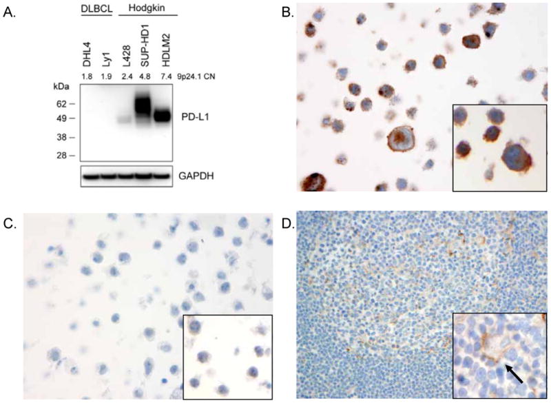
Purpose: Programmed cell death ligand 1 (PD-L1) is an immunomodulatory molecule expressed by antigen-presenting cells and select tumors that engages receptors on T cells to inhibit T-cell immunity. Immunotherapies targeting the PD-1/PD-L1 pathway have shown durable antitumor effects in a subset of patients with solid tumors. PD-L1 can be expressed by Reed-Sternberg cells comprising classical Hodgkin lymphoma (CHL) and by malignant B cells comprising EBV-positive posttransplant lymphoproliferative disorders (PTLD). We sought to determine whether the expression of PD-L1 represents a general strategy of immune evasion among aggressive B-cell lymphomas and virus- and immunodeficiency-associated tumors.

Experimental design: Using novel antibodies and formalin-fixed, paraffin-embedded (FFPE) tissue biopsies, we examined 237 primary tumors for expression of PD-L1.

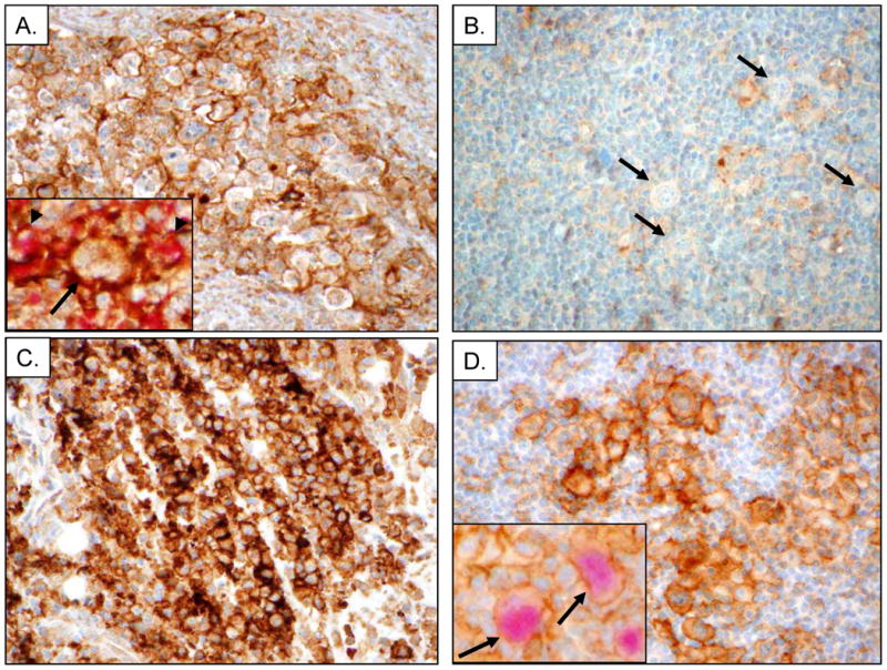
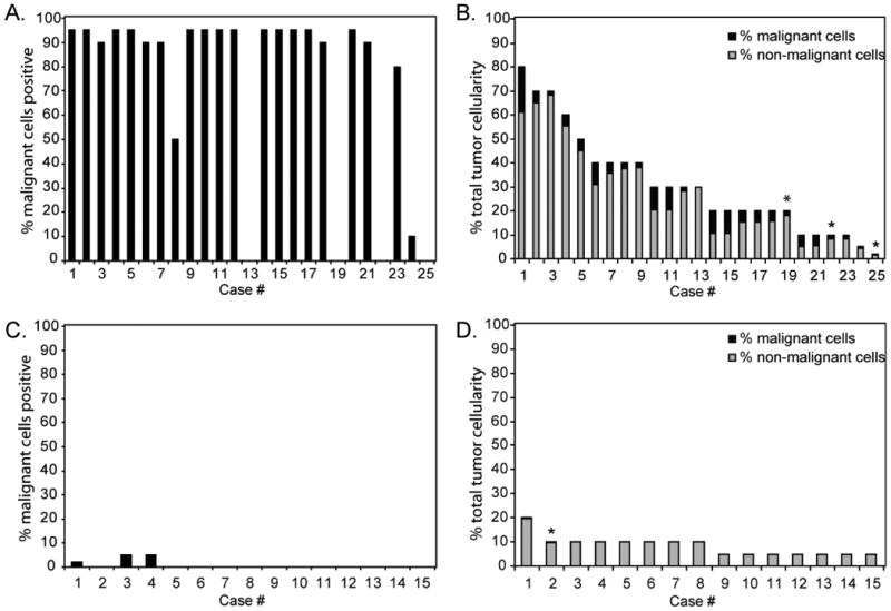
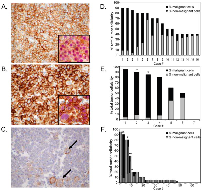
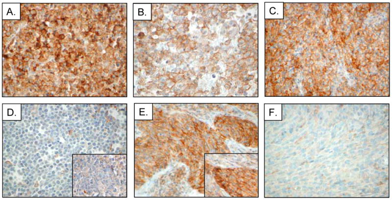
Results: Robust PD-L1 protein expression was found in the majority of nodular sclerosis and mixed cellularity CHL, primary mediastinal large B-cell lymphoma, T-cell/histiocyte-rich B-cell lymphoma, EBV-positive and -negative PTLD, and EBV-associated diffuse large B-cell lymphoma (DLBCL), plasmablastic lymphoma, extranodal NK/T-cell lymphoma, nasopharyngeal carcinoma, and HHV8-associated primary effusion lymphoma. Within these tumors, PD-L1 was highly expressed by malignant cells and tumor-infiltrating macrophages. In contrast, neither the malignant nor the nonmalignant cells comprising nodular lymphocyte-predominant Hodgkin lymphoma, DLBCL-not otherwise specified, Burkitt lymphoma, and HHV8-associated Kaposi sarcoma expressed detectable PD-L1.

Conclusion: Certain aggressive B-cell lymphomas and virus- and immunodeficiency-associated malignancies associated with an ineffective T-cell immune response express PD-L1 on tumor cells and infiltrating macrophages. These results identify a group of neoplasms that should be considered for PD-1/PD-L1-directed therapies, and validate methods to detect PD-L1 in FFPE tissue biopsies.


Conflict of interest statement

Conflicts of interest: GJF has patents and receives patent roylaties on the PD-1 pathway. There are no other conflicts of interest from the authors to disclose.

Figures

Figure 1
Figure 1
PD-L1 antibody validation. (A) Western blot analysis of cell lysates derived from genetically characterized diffuse large B-cell lymphoma (DLBCL) cell lines (DHL4, Ly1) and the Hodgkin lymphoma cell lines (L428, SUP-HD1, HDLM2). Lysates were probed with a rabbit monoclonal antibody recognizing PD-L1 (top panel) showing a band at the expected size of the fully glycosylated form of PD-L1 (∼55 kDa). The gene copy number (CN) for the CD274 (PD-L1) locus, 9p24.1, is shown, as reported previously, for each cell line. Equal loading was demonstrated by probing for GAPDH. IHC analysis of formalin-fixed, paraffin embedded (FFPE) Hodgkin cell line HDLM2 (B) and DLBCL cell line DHL4 (C) stained with the rabbit anti-PD-L1 antibody showing membranous staining of the HDLM2 cells but no staining of DHL4 cells. Insets show similar staining patterns with the mouse monoclonal anti-PD-L1 antibody. (D) FFPE human tonsil stained with rabbit anti-PD-L1 antibody showing little staining of lymphoid cells and weak membranous staining of occasional macrophages (inset, arrow).
Figure 2
Figure 2
Immunohistochemical analysis of PD-L1 in CHL, NLPHL, PMLBCL and TCRLBCL. (A) Representative example nodular sclerosis classical Hodgkin lymphoma (NSCHL) stained with the rabbit anti-PD-L1 antibody showing distinct membranous staining of Reed-Sternberg (RS) cells and intra-tumoral macrophages. Inset shows staining with PD-L1 (brown) highlighting the cell membrane of RS cells (arrow) as well as non-malignant cells, and macrophages (arrowheads) double stained with PD-L1 and the macrophage marker CD68 (red). (B) Representative case of nodular lymphocyte-predominant Hodgkin lymphoma (NLPHL) stained with rabbit anti-PD-L1 showing LP cells (arrows) that are negative for PD-L1. (C) Representative case of primary mediastinal large B-cell lymphoma (PMBCL) stained with rabbit anti-PD-L1 antibody showing predominantly membranous staining of lymphoma cells. (D) Representative case of T-cell/histiocyte-rich large B-cell lymphoma (TCHRBCL) stained with rabbit anti-PD-L1 antibody. Inset shows staining with PD-L1 (brown) highlighting macrophages adjacent to scattered malignant cells (arrow) that are double stained for PD-L1 and the B-cell lineage marker PAX5 (red).
Figure 3
Figure 3
Graphical representation of the percentage of the malignant cells (A, C) and the percentage of the total tumor cellularity (malignant and non-malignant cells) (B, D) staining positive for PD-L1 in each case of NSCHL (A, B) and NLPHL (C, D) examined. * indicates cases exhibiting predominantly cytoplasmic staining.
Figure 4
Figure 4
PD-L1 expression in EBV-positive DLBCLs, EBV-negative PTLD and DLBCL-NOS. (A) Representative case of an EBV-positive immunodeficiency-associated diffuse large B-cell lymphoma (DLBCL) stained with rabbit anti-PD-L1 showing distinct membranous staining of the lymphoma cells. Inset shows a case of EBV-positive DLBCL of the elderly double stained for PD-L1 (brown) and PAX5 (red). (B) Representative case of an EBV-negative post-transplant lymphoproliferative disorder (PTLD) stained with rabbit anti-PD-L1 showing distinct membranous staining of the lymphoma cells. Inset shows double staining of the same case for PD-L1 and PAX5. (C) Case of DLBCL-NOS stained with rabbit anti-PD-L1 showing no staining of the lymphoma cells but scattered intra-tumoral macrophages filled with apoptotic debris showing weak membrane positivity for PD-L1. (D-F) Graphical representation of the contribution of malignant and non-malignant cells staining positive for PD-L1 to total PD-L1 staining in each case of EBV+ DLBCL of the elderly/immunodeficiency-associated, EBV-negative PTLD, and DLBCL-NOS, respectively. * indicates cases exhibiting predominantly cytoplasmic staining.
Figure 5
Figure 5
Immunohistochemical analysis of PD-L1 expression in viral-associated lymphomas and additional cancers. Representative cases of (A) EBV-positive plasmablastic lymphoma (PBL), (B) HHV8-positive primary effusion lymphoma (PEL), (C) EBV-positive extranodal NK/T-cell lymphoma (ENKTCL), (D) EBV-positive Burkitt lymphoma (BL), (E) EBV-positive nasopharyngeal carcinoma (NPC), and (F) HHV8-positive Kaposi sarcoma (KS) stained with rabbit anti-PD-L1 or mouse anti-PD-L1 (insets).

Comment in

References

    1. Keir ME, Butte MJ, Freeman GJ, Sharpe AH. PD-1 and its ligands in tolerance and immunity. Annu Rev Immunol. 2008;26:677–704. - PMC - PubMed
    1. Freeman GJ, Long AJ, Iwai Y, Bourque K, Chernova T, Nishimura H, et al. Engagement of the PD-1 immunoinhibitory receptor by a novel B7 family member leads to negative regulation of lymphocyte activation. J Exp Med. 2000;192:1027–34. - PMC - PubMed
    1. Brahmer JR, Tykodi SS, Chow LQM, Hwu WJ, Topalian SL, Hwu P, et al. Safety and activity of anti-PD-L1 antibody in patients with advanced cancer. N Engl J Med. 2012;366:2455–65. - PMC - PubMed
    1. Topalian SL, Hodi FS, Brahmer JR, Gettinger SN, Smith DC, McDermott DF, et al. Safety, activity, and immune correlates of anti-PD-1 antibody in cancer. N Engl J Med. 2012;366:2443–54. - PMC - PubMed
    1. Green MR, Monti S, Rodig SJ, Juszczynski P, Currie T, O'Donnell E, et al. Integrative analysis reveals selective 9p24.1 amplification, increased PD-1 ligand expression, and further induction via JAK2 in nodular sclerosing Hodgkin lymphoma and primary mediastinal large B-cell lymphoma. Blood. 2010;116:3268–77. - PMC - PubMed

Publication types

MeSH terms