

Functional MRI of cerebellar activity during eyeblink classical conditioning in children and adults
- PMID: 23674498
- PMCID: PMC3823743
- DOI: 10.1002/hbm.22261
Functional MRI of cerebellar activity during eyeblink classical conditioning in children and adults
Abstract
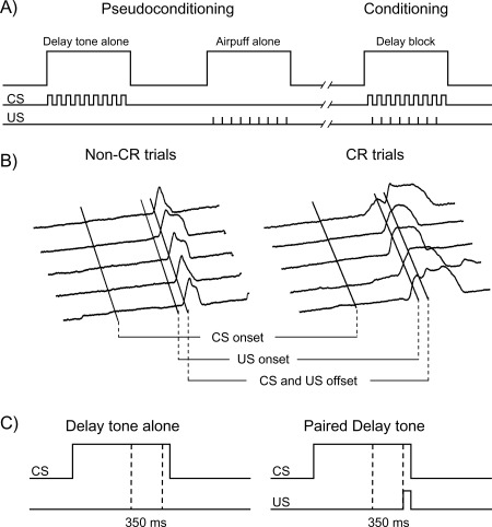
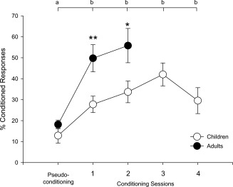
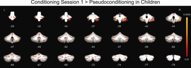
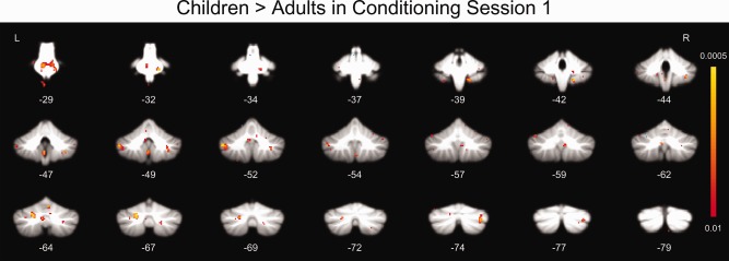
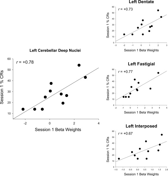
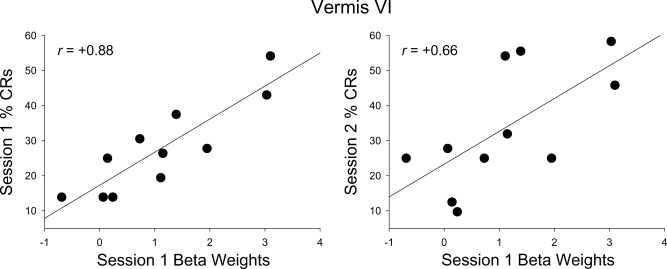
This study characterized human cerebellar activity during eyeblink classical conditioning (EBC) in children and adults using functional magnetic resonance imaging (fMRI). During fMRI, participants were administered delay conditioning trials, in which the conditioned stimulus (a tone) precedes, overlaps, and coterminates with the unconditioned stimulus (a corneal airpuff). Behavioral eyeblink responses and brain activation were measured concurrently during two phases: pseudoconditioning, involving presentations of tone alone and airpuff alone, and conditioning, during which the tone and airpuff were paired. Although all participants demonstrated significant conditioning, the adults produced more conditioned responses (CRs) than the children. When brain activations during pseudoconditioning were subtracted from those elicited during conditioning, significant activity was distributed throughout the cerebellar cortex (Crus I-II, lateral lobules IV-IX, and vermis IV-VI) in all participants, suggesting multiple sites of associative learning-related plasticity. Despite their less optimal behavioral performance, the children showed greater responding in the pons, lateral lobules VIII, IX, and Crus I, and vermis VI, suggesting that they may require greater activation and/or the recruitment of supplementary structures to achieve successful conditioning. Correlation analyses relating brain activations to behavioral CRs showed a positive association of activity in cerebellar deep nuclei (including dentate, fastigial, and interposed nuclei) and vermis VI with CRs in the children. This is the first study to compare cerebellar cortical and deep nuclei activations in children versus adults during EBC.
Keywords: cerebellum; development; learning; memory; neuroimaging.
Copyright © 2013 Wiley Periodicals, Inc.
Figures
References
-
- Amunts K, Kedo O, Kindler M, Pieperhoff P, Mohlberg H, Shah NJ, Habel U, Schneider F, Zilles K (2005): Cytoarchitectonic mapping of the human amygdala, hippocampal region and entorhinal cortex: Intersubject variability and probability maps. Anat Embryol (Berl),210:343–352. - PubMed
-
- Berger TW, Alger B, Thompson RF (1976): Neuronal substrate of classical conditioning in the hippocampus. Science 192:483–485. - PubMed
-
- Bracha V, Webster ML, Winters NK, Irwin KB, Bloedel JR (1994): Effects of muscimol inactivation of the cerebellar interposed‐dentate nuclear complex on the performance of the nictitating membrane response in the rabbit. Exp Brain Res 100:453–468. - PubMed
-
- Brown KL, Pagani JH, Stanton ME (2006): The ontogeny of interstimulus interval (ISI): Discrimination of the conditioned eyeblink response in rats. Behav Neurosci 120:1057–1070. - PubMed
Publication types
MeSH terms
Grants and funding
LinkOut - more resources
Full Text Sources
Other Literature Sources
