. 2013 May 28;110(22):E2038-45.
doi: 10.1073/pnas.1305716110. Epub 2013 May 14.

Direct interaction between the TnsA and TnsB subunits controls the heteromeric Tn7 transposase

Affiliations

Direct interaction between the TnsA and TnsB subunits controls the heteromeric Tn7 transposase

Ki Young Choi et al. Proc Natl Acad Sci U S A. .

Abstract

The transposon Tn7 transposase that recognizes the transposon ends and mediates breakage and joining is heteromeric. It contains the Tn7-encoded proteins TnsB, which binds specifically to the transposon ends and carries out breakage and joining at the 3' ends, and TnsA, which carries out breakage at the 5' ends of Tn7. TnsA apparently does not bind specifically to DNA, and we have hypothesized that it is recruited to the ends by interaction with TnsB. In this work, we show that TnsA and TnsB interact directly and identify several TnsA and TnsB amino acids involved in this interaction. We also show that TnsA can stimulate two key activities of TnsB, specific binding to the ends and pairing of the Tn7 ends. The ends of Tn7 are structurally asymmetric (i.e., contain different numbers of TnsB-binding sites), and Tn7 also is functionally asymmetric, inserting into its specific target site, attachment site attTn7 (attTn7) in a single orientation. Moreover, Tn7 elements containing two Tn7 right ends can transpose, but elements with two Tn7 left ends cannot. We show here that TnsA + TnsB are unable to pair the ends of a Tn7 element containing two Tn7 left ends. This pairing defect likely contributes to the inability of Tn7 elements with two Tn7 left ends to transpose.

Keywords: asymmetric transposon ends; protein-DNA interaction; protein–protein interaction; transposition.


Conflict of interest statement

The authors declare no conflict of interest.

Figures
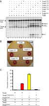
Fig. 1.
Fig. 1.
In vivo and in vitro comparisons of TnsA gain-of-function mutants. (A) In vitro Tn7 transposition assays with TnsA gain-of-function mutants. Reactions were carried out in high glycerol (20%, vol/vol) and 15 mM Mg2+, digested with a restriction enzyme that cleaved once in the donor backbone. DNA DSBs at the right or left end of Tn7 result in DSB.R and DSB.L, respectively. DSBs at both ends result in an ELT. M, size markers. (B) Comparison of TnsA gain-of-function mutants by papillation in cells containing TnsA mutants and wild-type TnsB. Cell suspensions as indicated were spotted on MacConkey lactose plates. (C) In vivo lambda hop assay with Tns mutants. The frequency of the transposition of a miniTn7Kan element from an integration-defective lambda phage into the E. coli chromosome was measured in strains containing the various tnsA, tnsB, and tnsC genes. The results are the average of three independent experiments and are shown as the number of transposition events.
Fig. 2.
Fig. 2.
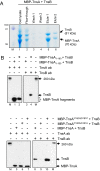
TnsA interacts directly with TnsB. (A) The elution profiles of MBP–TnsA and TnsB from an amylose column. Fractions were displayed on an SDS/PAGE gel and stained with Coomassie blue. A single gel is shown. M, size markers; lane 1, crude extract; lane 2, flow-through fraction; lanes 3 and 4, wash fractions; lanes 5 and 6, fractions eluted with maltose buffer. (B) Western blot of maltose-eluted fractions using extracts containing MBP–TnsA mutated proteins with TnsA or TnsB antibody as indicated. M, size markers; lanes 1 and 3, maltose-eluted fraction using MBP–TnsA1–160 blotted with TnsA and TnsB antibodies; lanes 2 and 4, maltose-eluted fraction using MBP–TnsA1–190 blotted with TnsA and TnsB; lanes 5 and 8, maltose-eluted fraction using TnsA blotted with TnsA and TnsB antibodies; lanes 6 and 9, maltose-eluted fraction using TnsAY180A/S181A blotted with TnsA and TnsB antibodies; lanes 7 and 10, maltose-eluted fraction using TnsAY180H/S181P blotted with TnsA and TnsB antibodies. Equivalent reactions were run on the same gel, and different lanes were blotted with either anti-TnsA or anti-TnsB antibody.
Fig. 3.
Fig. 3.
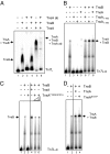
TnsA stimulates TnsB binding at the end of Tn7. The binding of TnsB to Tn7L1–166 and Tn7L1–30 is evaluated by gel retardation assays. (A) TnsB–Tn7L can be supershifted with TnsA and an antibody against TnsA. TnsA makes a new complex from the TnsB–Tn7L end complex. Lane 1, no protein; lane 2, TnsB; lane 3, TnsA + anti-TnsA antibody; lane 4, TnsB + anti-TnsA antibody; lane 5, TnsB + TnsA; lane 6, TnsB+TnsA and anti-TnsA antibody. (B) TnsA stimulation of TnsB binding requires TnsA161–190. Lane 1, no protein; lane 2, TnsB; lane 3, TnsA1–160; lane 4, TnsA1–190; lane 5, TnsA; lane 6, TnsB + TnsA1–160; lane 7, TnsB + TnsA1–190; lane 8, TnsB + TnsA. (C) TnsAY180A/S181A does not stimulate TnsB binding. Lane 1, no protein; lane 2. TnsB; lane 3, TnsAY180A/S181A; lane 4, TnsB + TnsA; lane 5, TnsB+TnsAY180A/S181A; lane 6, TnsB+ 3× TnsAY180A/S181A. As indicated by the vertical line, different sections from the same gel have been juxtaposed next to each other. (D) The gain-of-function mutant TnsAS181P stimulates TnsB binding more than TnsA. Lane 1, no protein; lane 2, TnsB; lane 3, TnsB + TnsA; lane 4, TnsB + TnsAS181P. As indicated by the vertical line in C and D, different sections from the same gel have been juxtaposed next to each other.
Fig. 4.
Fig. 4.
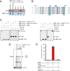
Interaction of TnsB mutants with TnsA. (A) Copurification of TnsB401–480 with MBP–TnsA. The copurification of TnsB401–480 with MBP–TnsA after elution from an amylose column with maltose was evaluated by SDS/PAGE, followed by Western blotting with TnsB antibody and staining with Coomassie blue. Lane 1, cell lysate; lane 2, flow-through fraction; lane 3, wash fraction; lanes 4, 5, and 6, fractions eluted with maltose. Equivalent reactions were run on the same gel, and different lanes were blotted with either anti-TnsA or anti-TnsB antibody. (B) Multiple sequence alignment of TnsBs between amino acids 440 and 480 produced using the software T-Coffee (www.ebi.ac.uk/Tools/msa/tcoffee/) (38). B, E. coli (NP_065319) (TnsB in this work); 1, Shewanella putrefaciens CN-32 (YP_001185440); 2, Tolumonas auensis DSM 9187 (YP_002891520); 3, Acinetobacter baumannii ATCC17978 (ABO12968); 4, Idiomarina loihiensis L2TR (YP_156993). Conserved positions are highlighted in blue. Changes at positions indicated by blue arrows lead to loss of TnsB interaction with TnsA. Changes at underscored positions did not affect interaction with TnsA. (C) TnsBD467A/L468A does not interact with MBP–TnsA. Analysis of the copurification of TnsB truncations with MBP–TnsA after elution from an amylose column with maltose was evaluated by SDS/PAGE, followed by Western blotting with anti-TnsA and anti-TnsB antibodies as indicated. M, size markers; lanes 1, 4, 7, and 9, MBP–TnsA + TnsB; lanes 2 and 5, MBP–TnsA + TnsBY458A/D459A/R460A; lanes 3 and 6, MBP–TnsA + TnsBW478A/G479A; lanes 8 and 10, MBP–TnsA + TnsBD467A/L468A. Equivalent reactions were run on the same gel, and different lanes were blotted with either anti-TnsA or anti-TnsB antibody. (D) TnsBD467A/L468A binding to a Tn7 end is not stimulated by TnsA. TnsB binding to Tn7L-α was evaluated by EMSA with different concentrations of TnsA on the same gel. Lane 1, no TnsB; lane 2, TnsBD467A/L468A; lane 3, TnsBD467A/L468A + 1× TnsA; lane 4, TnsBD467A/L468A + 3× TnsA; lane 5, TnsBD467A/L468A + 9× TnsA. As indicated by the vertical line, different sections from the same gel have been juxtaposed next to each other. (E) TnsBD467A/L468A is inactive in transposition in vivo. Transposition in the presence of TnsA, TnsB, and TnsC was measured in a lambda hop assay. The results are the average of three independent experiments and are shown as the number of transposition events.
Fig. 5.
Fig. 5.
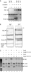
In vitro formation of the PEC by TnsB and TnsA. (A) The formation of a PEC is examined under various conditions as indicated. (B) The stability of the PEC is different in Mg2+ and 5% (vol/vol) glycerol vs. Mn2+ and 20% (vol/vol) glycerol. The stability over time of a PEC formed in the presence of either Mg2+ and 5% (vol/vol) glycerol (lanes 1–5) or Mn2+ and 20% (vol/vol) glycerol (lanes 6–10) is examined after the addition of excess TnsB-binding sites. (C) TnsA stimulates TnsB to form the PEC. M, size markers; lane 1, no protein addition; lane 2, TnsB, lane 3, TnsA; lane 4, TnsB + TnsA; lane 5. loss-of-function TnsAY180A/S181A; lane 6, TnsB + loss-of-function TnsAY180A/S181A; lane 7, loss-of-function TnsBD467A/L468A; lane 8, loss-of-function TnsBD467A/L468A + TnsA.
Fig. 6.
Fig. 6.
Formation of PEC with various Tn7 elements. End pairing of Tn7L–Tn7R, Tn7L–Tn7L, and Tn7R–Tn7R donor plasmids was analyzed in the presence by TnsB and TnsA as indicated. Lanes 1–4, analysis of Tn7L–Tn7R; lanes 5–8, analysis of Tn7L–Tn7L; lanes 9–1, analysis of Tn7R–Tn7R.

Similar articles

Cited by

References

    1. Craig NL, Craigie R, Gellert M, Lambowitz A. Mobile DNA II. Washington, DC: ASM; 2002.
    1. Peters JE, Craig NL. Tn7: Smarter than we thought. Nat Rev Mol Cell Biol. 2001;2(11):806–814. - PubMed
    1. Li Z, Craig NL, Peters JE. 2013. in Bacterial Integrative Mobile Genetic Elements, eds Roberts A, Mullany P (Landes Bioscience, London), pp. 1–32.
    1. May EW, Craig NL. Switching from cut-and-paste to replicative Tn7 transposition. Science. 1996;272(5260):401–404. - PubMed
    1. Sarnovsky RJ, May EW, Craig NL. The Tn7 transposase is a heteromeric complex in which DNA breakage and joining activities are distributed between different gene products. EMBO J. 1996;15(22):6348–6361. - PMC - PubMed

Publication types

MeSH terms

LinkOut - more resources