

The role of lithium carbonate and lithium citrate in regulating urinary citrate level and preventing nephrolithiasis
- PMID: 23675140
- PMCID: PMC3614779
The role of lithium carbonate and lithium citrate in regulating urinary citrate level and preventing nephrolithiasis
Abstract
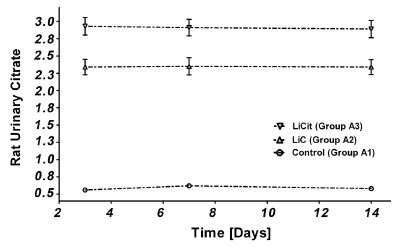
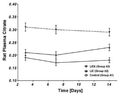
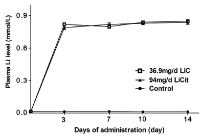
Background and purpose: Urinary Citrate is an inhibitor of Calcium oxalate stone formation. It is reabsorbed in the proximal kidney through sodium dicarboxylate co-transporters (NaDC-1, NaDC-3) present in the renal tubular epithelium. Lithium (Li) is a known potent inhibitor of these transporters. We investigated the effect of lithium carbonate (LiC) and lithium citrate (LiCit) in regulating urinary citrate levels and preventing nephrolithiasis (NL) in the rat model.
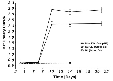
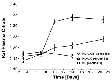
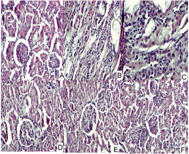
Experimental approach: We took 220 Wistar rats and induced nephrolithiasis in 130 of them by administering high doses of 5% ammonium oxalate (AmOx) for seven days and labeled them as Group B. Rest were labeled as Group A. Each group was then divided into 3 subgroups. First sub-group acted as control while other two were treated with either lithium carbonate (LiC) or lithium citrate (LiCit) for 21 days. Ten rats from each of the six sub-groups were randomly selected for sacrifice on 3(rd), 7(th) and 14(th) day and additional 10(th) and 21(st) day from Li treated groups. Blood and urine samples were collected and analyzed on these days. The kidneys of the sacrificed rats were dissected and studied under light microscopy for crystal deposition (left kidney) and histological changes (right kidney).
Key results: Urinary citrate levels were significantly increased in response to either LiC (p<0.001) or LiCit (p<0.001). Increased urinary citrate levels resulted in the reduction of calcium oxalate (CaOx) crystal deposition, kidney tubular dilatation and infiltration of inflammatory cell in the tubulo-interstitium.
Conclusions and implications: Use of lithium salts might be a potentially useful approach in the prevention of recurrent NL.
Keywords: kidney calculi; lithium; nephrolithiasis; urinary citrate levels; urolithiasis.
Figures
Similar articles
-
Sodium dicarboxylate cotransporter-1 expression in renal tissues and its role in rat experimental nephrolithiasis.J Nephrol. 2004 Jan-Feb;17(1):34-42. J Nephrol. 2004. PMID: 15151257
-
rs11567842 SNP in SLC13A2 gene associates with hypocitraturia in Thai patients with nephrolithiasis.Genes Genomics. 2018 Sep;40(9):965-972. doi: 10.1007/s13258-018-0702-4. Epub 2018 May 17. Genes Genomics. 2018. PMID: 30155711
-
Dietary treatment of urinary risk factors for renal stone formation. A review of CLU Working Group.Arch Ital Urol Androl. 2015 Jul 7;87(2):105-20. doi: 10.4081/aiua.2015.2.105. Arch Ital Urol Androl. 2015. PMID: 26150027 Review.
-
Small intestine resection increases oxalate and citrate transporter expression and calcium oxalate crystal formation in rat hyperoxaluric kidneys.Clin Sci (Lond). 2020 Oct 16;134(19):2565-2580. doi: 10.1042/CS20200973. Clin Sci (Lond). 2020. PMID: 33006369 Free PMC article.
-
Urinary citrate and renal stone disease: the preventive role of alkali citrate treatment.Arch Ital Urol Androl. 2009 Sep;81(3):182-7. Arch Ital Urol Androl. 2009. PMID: 19911682 Review.
Cited by
-
Protective potential of Angelica sinensis polysaccharide extract against ethylene glycol-induced calcium oxalate urolithiasis.Ren Fail. 2018 Nov;40(1):618-627. doi: 10.1080/0886022X.2018.1496935. Ren Fail. 2018. PMID: 30396308 Free PMC article.
-
Homeostasis of chosen bioelements in organs of rats receiving lithium and/or selenium.Biometals. 2016 Oct;29(5):873-9. doi: 10.1007/s10534-016-9958-9. Epub 2016 Jul 30. Biometals. 2016. PMID: 27476158 Free PMC article.
-
An Explanation of the Underlying Mechanisms for the In Vitro and In Vivo Antiurolithic Activity of Glechoma longituba.Oxid Med Cell Longev. 2016;2016:3134919. doi: 10.1155/2016/3134919. Epub 2016 Oct 20. Oxid Med Cell Longev. 2016. PMID: 27840669 Free PMC article.
-
The elementome of calcium-based urinary stones and its role in urolithiasis.Nat Rev Urol. 2015 Oct;12(10):543-57. doi: 10.1038/nrurol.2015.208. Epub 2015 Sep 1. Nat Rev Urol. 2015. PMID: 26334088 Free PMC article. Review.
References
-
- Meyer JL, Smith LH. Growth of calcium oxalate crystals. II. Inhibition by natural urinary crystal growth inhibitors. Invest Urol. 1975;13(1):36–39. - PubMed
-
- Kok DJ, Papapoulos SE, Bijvoet OL. Excessive crystal agglomeration with low citrate excretion in recurrent stone-formers. Lancet. 1986;1(8489):1056–1058. - PubMed
-
- Nicar MJ, Hill K, Pak CY. Inhibition by citrate of spontaneous precipitation of calcium oxalate in vitro . J. Bone Miner Res. 1987;2(3):215–220. - PubMed
-
- Menon M, Mahle CJ. Urinary citrate excretion in patients with renal calculi. J. Urol. 1983;129(6):1158–1160. - PubMed
-
- Levy FL, Adams-Huet B, Pak CY. Ambulatory evaluation of nephrolithiasis: an update of a 1980 protocol. Am. J. Med. 1995;98(1):50–59. - PubMed
LinkOut - more resources
Full Text Sources
Miscellaneous