. 2009 Dec;5(4):411-20.

Statins inhibit the proliferation and induce cell death of human papilloma virus positive and negative cervical cancer cells

Affiliations

Statins inhibit the proliferation and induce cell death of human papilloma virus positive and negative cervical cancer cells

María Elena Crescencio et al. Int J Biomed Sci. 2009 Dec.

Abstract

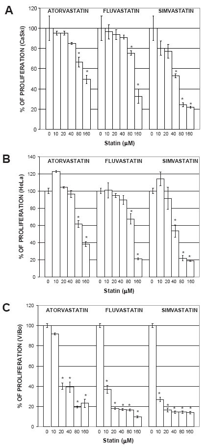
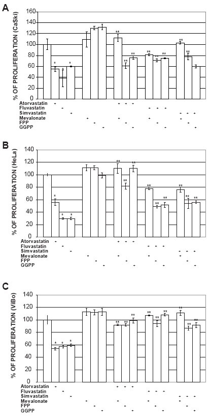
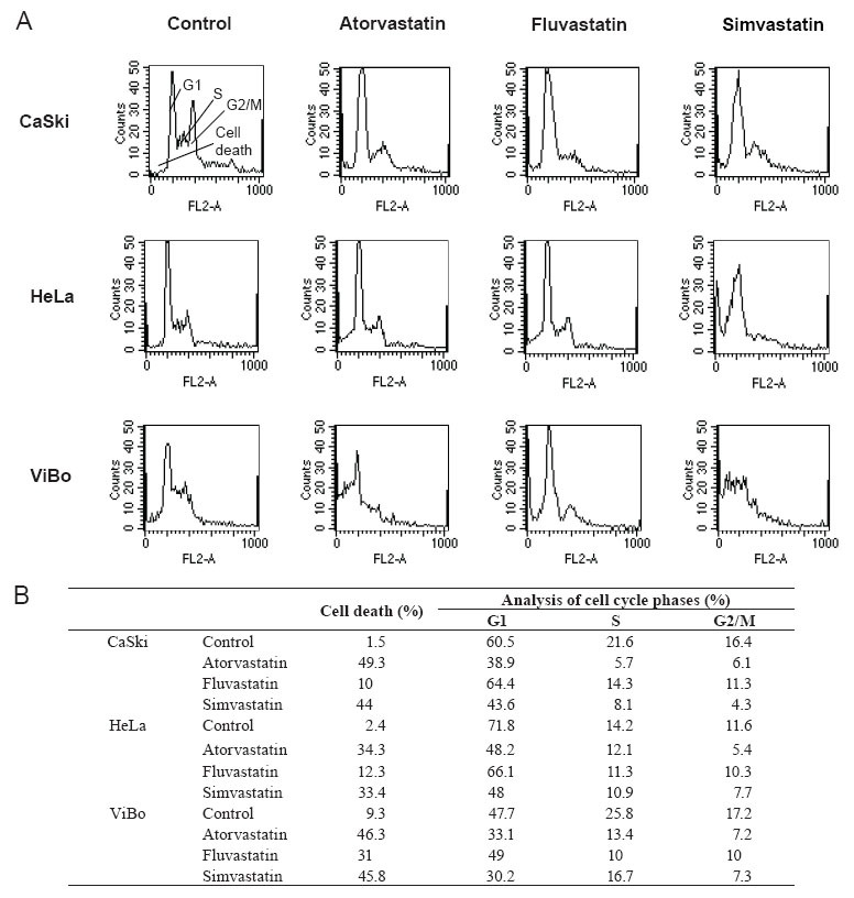
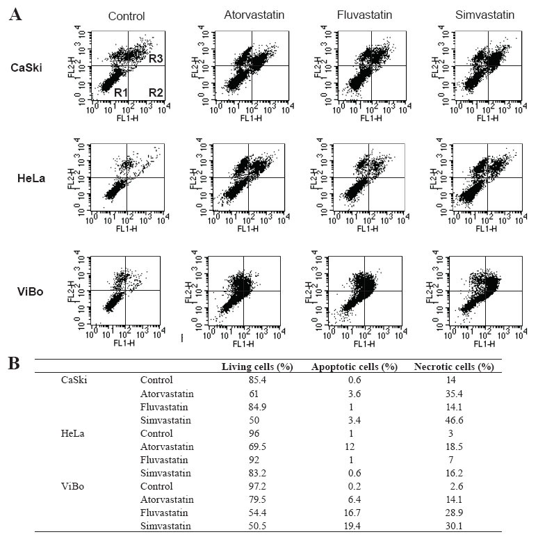
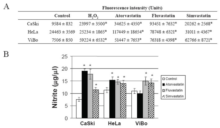
Statins, competitive inhibitors of 3-hydroxy-3-methylglutaryl-coenzyme A (HMG-CoA) reductase, have anti-tumoral effects on multiple cancer types; however, little is known about their effect on cervical cancer. We evaluated the effect on proliferation, cell cycle, oxidative stress and cell death of three statins on CaSki, HeLa (HPV(+)) and ViBo (HPV(-)) cervical cancer cell lines. Cell proliferation was assayed by crystal violet staining, cell cycle by flow cytometry and cell death by annexin-V staining. Reactive oxygen species (ROS) production was evaluated by the oxidation of 2,7-dichlorofluorescein diacetate and nitrite concentration (an indirect measure of nitric oxide (NO) production), by the Griess reaction. Inhibition of cell proliferation by atorvastatin, fluvastatin and simvastatin was dose-dependent. ViBo cells were the most responsive. Statins did not affect the cell cycle, instead they induced cell death. The antiproliferative effect in ViBo cells was completely inhibited with mevalonate, farnesyl pyrophosphate (FPP) and geranylgeranyl pyrophosphate (GGPP) treatments. In contrast, cell proliferation of CaSki and HeLa cells was partially (33%) rescued with these intermediates. The three statins increased ROS and nitrite production, mainly in the ViBo cell line. These results suggest that statins exert anti-tumoral effects on cervical cancer through inhibition of cell proliferation and induction of cell death and oxidative stress. Statins could be an aid in the treatment of cervical cancer, especially in HPV(-) tumors.

Keywords: cell cycle; cell death; cervical cancer; human papilloma virus; mevalonate pathway; oxidative stress; proliferation; statins.


Figures

Figure 1
Figure 1
Effect of statins on cell proliferation. CaSki (A), HeLa (B) and ViBo cells (C) were treated with 0, 10, 20, 40, 80 and 160 μM atorvastatin, fluvastatin or simvastatin for 48 h. Cell proliferation was evaluated by crystal violet staining according to materials and methods. * Indicates a p<0.01 compared to control cells.
Figure 2
Figure 2
Effect of mevalonate pathway intermediates on the inhibition of proliferation induced by statins. Cells were cultured with mevalonate (100 μM), farnesyl pyrophosphate (FPP) (20 μM) or geranylgeranyl pyrophosphate (GGPP) (20 μM) 2 h before statin treatment and cell proliferation was evaluated by crystal violet staining after 48 h. A) CaSki cells, B) HeLa cells and C) ViBo cells. The results are expressed as percentage of proliferation with respect to untreated cells and are showed as mean ± SD of one experiment (n=5). The results correspond to a representative experiment of three independent assays. * Indicates a p<0.01 compared with control cells and ** a p<0.01 compared with statin-treated cells.
Figure 3
Figure 3
Effect of statins on the phases of the cell cycle. Cells were cultured without (Control) and with atorvastatin, fluvastatin or simvastatin and the percentage of cells in each phase of the cell cycle was evaluated by flow cytometry 48 h after. A) shows the histograms and B) shows the percentage of cells in each phase of the cell cycle obtained with the Modift software (Becton Dickinson). Upper left histogram shows the regions containing the cells in all the phases of the cell cycle (G1, S, G2/M) and cell death. The results correspond to a representative experiment of three independent assays.
Figure 4
Figure 4
Effect of statins on cell death evaluated by annexin-V-fluos. Cells were cultured without (Control) and with statins for 48 h. A) Cells were stained with annexin-V-fluos (FL1-H) and propidium iodide (PI) (FL2-H). Lower left panel shows living cells (R1), lower right panel, apoptotic cells (R2), and upper right panel, necrotic cells (R3). B) Graphic representation of the percentage of cells in each panel obtained by flow cytometry with the Modift software. The results correspond to a representative experiment of three independent assays.
Figure 5
Figure 5
ROS and nitrite production by statins-treated cell lines. A) ROS production was evaluated using DCFDA and flow cytometry. The results are expressed as fluorescence intensity and are showed as mean ± SD of one experiment (n=5). Fluorescence intensity (units) was calculated by multiplying the number of events by the mean fluorescence intensity value in a region selected manually with positive fluorescence in the channel FL-1 (DCF). The results correspond to a representative experiment of three independent assays. Cells without DCFDA were used as a negative control. B) Nitrite concentration was determined by Griess reagent reactivity (see methods). The results are expressed as mean ± SD of one experiment (n=5). We show a representative experiment of three independent assays. *Indicates a p < 0.01 in comparison with the control (cells without treatment).

Similar articles

Cited by

References

    1. Jakobisiak M, Golab J. Potential antitumor effects of statins. Int. J. Oncol. 2003;23:1055. - PubMed
    1. Goldstein JL, Brown MS. Regulation of the mevalonate pathway. Nature. 1990;343:425. - PubMed
    1. Alegret M, Silvestre JS. Pleiotropic effects of statins and related pharmacological experimental approaches. Methods Find Exp. Clin. Pharmacol. 2006;28:627. - PubMed
    1. Graaf MR, Richel DJ, van Noorden CJ, GucHeLaar HJ. Effects of statins and farnesyltransferase inhibitors on the development and progression of cancer. Cancer Treat. Rev. 2004;30:609. - PubMed
    1. Dimitroulakos J, Ye LY, Benzaquen M, Moore MJ, et al. Differential sensitivity of various pediatric cancers and squamous cell carcinomas to lovastatin-induced apoptosis: therapeutic implications. Clin. Cancer Res. 2001;7:158. - PubMed

LinkOut - more resources