. 2010 Jun;6(2):111-9.

Diethylcarbamazine and non-diethylcarbamazine related bancroftian granuloma: an immunohistochemical study of eosinophil toxic proteins

Affiliations

Diethylcarbamazine and non-diethylcarbamazine related bancroftian granuloma: an immunohistochemical study of eosinophil toxic proteins

Jose Figueredo-Silva et al. Int J Biomed Sci. 2010 Jun.

Abstract

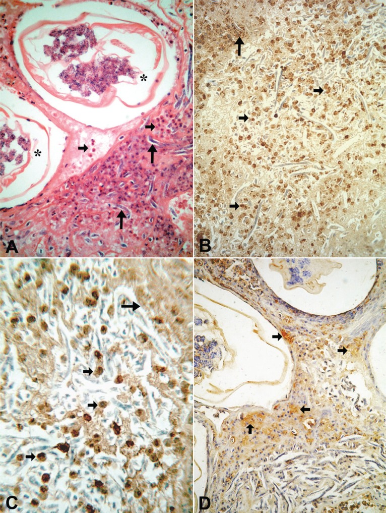
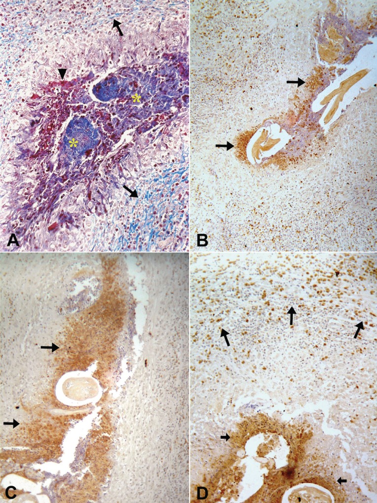
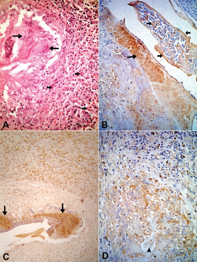
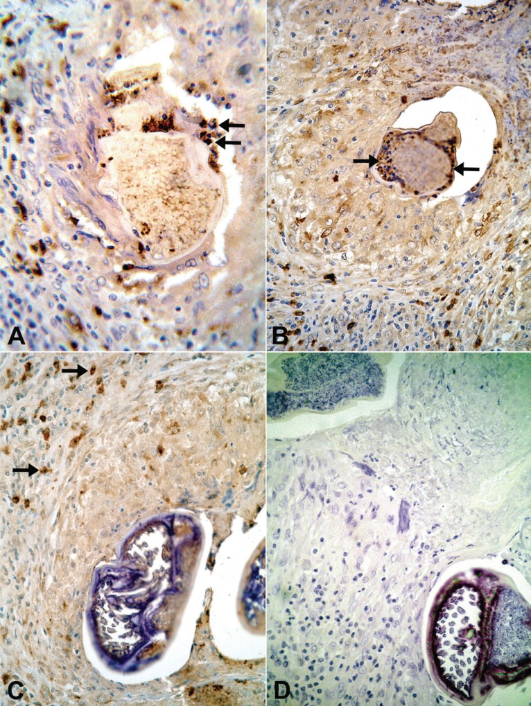
It has been suggested, mostly using in vitro experiments, that defenses against parasites involve mainly activated eosinophils and their toxic proteins, such as major basic protein (MBP), eosinophil cationic protein (ECP) and eosinophil peroxidase (EPO). Eosinophil degranulation has been described around degenerating onchocercal microfilariae in patients treated with diethylcarbamazine (DEC). In bancroftian filariasis, traditional histopathologic studies have shown remarkable numbers of eosinophils in granulomatous lesions associated with both DEC-induced and spontaneous death of adult Wuchereria bancrofti parasites. No immunohistochemical study targeting eosinophil degranulation has been previously performed in these granulomas, which are found mainly within intrascrotal lymphatic vessels. This investigation was undertaken in 22 (12 DEC-treated and 10 untreated) male patients in order to determine the immunohistochemical expressions of MBP, EPO and ECP in bancofitian granulomas, using the indirect method. Stained intact esosinophils, as well as granular, extra-cellular material positive for all three proteins, were found in all granulomas. The immunohistochemical patterns were similar in both DEC-treated and untreated cases, irrespective of microfilaremia, blood eosinophilia, and granuloma age. Positive intact cells were observed mostly at the periphery of the granulomas, whereas granular material predominated in central areas around dead or degenerating parasites. These results indicate that eosinophils accumulate in the granulomas and degranulate preferentially in close proximity to degenerating or dead adult parasites. In bancroftian granulomas, influx and degranulation of eosinophils are considered a consequence of parasite death, rather than its cause.

Keywords: Wuchereria bancrofti; bancroftian filariasis; eosinophil cationic protein; eosinophil peroxidase; eosinophils; filarial granuloma; immunohistochemistry; lymphatic fiariasis; major basic protein.


Figures

Figure 1
Figure 1
Twenty-one year-old microfilaremic (243 Mf/mL) treated patient (diethylcarbamazine 6mg/kg/day/12days) with a nodule obtained 7 days after the beginning DEC. (A) Transverse sections of degenerating adult worm (*) enclosed by fibrin-like material containing free Mf (large arrows) and inflammatory cells - mainly eosinophils (arrows) - without a granulomatous pattern (H & E. 400×). (B) Positive immunostaining for major basic protein showing large numbers of intact eosinophils (small arrows) and extracellular granular material surrounding degenerating free Mf (arrow) (400×). (C) The same immune pattern - intact cells (small arrows) and granular material (arrow) - is observed also for eosinophilic cationic protein, 600×. (D) Granular material (arrows) can be easily recognized as well as with eosinophil peroxidase staining, 400×.
Figure 2
Figure 2
Twenty-five year-old microfilaremic (1,350 Mf/mL) untreated patient with a nodule with unknown time of evolution. (A) A granulomatous inflammatory reaction around desintegrating adult worms (*) is observed; a rim of collagen deposition is seen at the periphery (arrows). Necrotic area is seen around the degenerated worms (arrow head). Manson trichome, 400x. Granular, extracellular positive material for major basic protein (B), eosinophil cationic protein (C) is condensed around dead adult worms (arrows) (100×). (D) Granular material stained for eosinophil peroxidase (small arrows) is also detected around the worms; intact marked cells predominate at periphery of the granuloma (arrows), 400×.
Figure 3
Figure 3
Nineteen year-old microfilaremic (124 Mf/mL) treated patient (diethylcarbamazine 6mg/kg single dose) with a nodule obtained 90 days after treatment. (A) A granulomatous response rich in eosinophils (small arrows) is observed around barely visible remnants of adult worm; the altered cuticle is seen as an eosinophilic line circumscribed by giant cells (arrows). H & E, 400×. (B) A tangential section of an adult female worm with abundant granular material positive for major basic protein (large arrow); the same material is also covering the cuticle and among intrauterine microfilariae (arrows), 400×. (C) Positive reaction is observed for eosinophilic cationic protein around degenerated worm (arrows), 100×. (D). Granular, extra-cellular material stained for eosinophilic peroxidase is present surrounding the remnants of parasite (arrow head), while intact brown, marked cells are detected at the periphery of the granuloma, 400×.
Figure 4
Figure 4
(A) Twenty-one year-old amicrofilaremic treated patient (diethylcarbamazine 6mg/kg/day/12 days) with a nodule obtained 10 days after beginning treatment. Intact cells marked for major basic protein are attached on parasite surface (arrows), 600×. (B, C and D) Nineteen year-old microfilaremic (1400 Mf/mL) untreated patient with a nodule with unknown time of evolution. (B) A collar of intact cells marked for eosinophilic peroxidase is observed on parasite surface (arrows), 400x. (C) Intact cells stained for eosinophilic cationic protein are present at the periphery of the granuloma (arrows); no positive cell is attached to the calcified wall of the parasite; only little residual amount of brown granular positive material is seen inside the parasite, 400×. (D) A control slide showed no immune staining for major basic protein. (Harris’ hematoxilyn, 400×).

References

    1. Rothenberg ME. Eosinophilia. N. Engl. J. Med. 1998;338(22):1592. - PubMed
    1. Klion AD, Nutman TB. The role of eosinophils in host defense against helminth parasites. J. Allergy Clin. Immunol. 2004;113(1):30. - PubMed
    1. Meeusen EN, Balic A. Do eosinophils have a role in the killing of helminth parasites? Parasitol Today. 2000;16(3):95. - PubMed
    1. Butterworth AE, Sturrock R, Houba V, et al. Eosinophils as mediators of antibody-dependent damage to schistosomula. Nature. 1975;256(5520):727. - PubMed
    1. Greene BM, Taylor HR, Aikawa M. Cellular killing of microfilariae of Onchocerca volvulus: eosinophil and neutrophil mediated immune serum dependent destruction. J. Immunol. 1981;127(4):1611. - PubMed

LinkOut - more resources