Interleukin-2 Functions in Anaplastic Large Cell Lymphoma Cells through Augmentation of Extracellular Signal-Regulated Kinases 1/2 Activation
- PMID: 23675235
- PMCID: PMC3614832
Interleukin-2 Functions in Anaplastic Large Cell Lymphoma Cells through Augmentation of Extracellular Signal-Regulated Kinases 1/2 Activation
Abstract
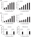
In addition to intrinsic genetic alterations, the effects of the extrinsic microenvironment also play a pathological role in cancer development. Altered chemokine/cytokine networks in the tumor microenvironment may contribute to the dysregulation of cellular functions in cancer cells. Anaplastic large cell lymphoma (ALCL) is an aggressive T-cell lymphoma caused by abnormal expression of anaplastic lymphoma kinase due to a chromosomal translocation. Notably, ALCL cells are also characterized by high-level expression of the high-affinity IL-2 receptor subunit CD25 on the cell surface. However, whether the IL-2/IL-2 receptor functions in ALCL cells and how this signaling affects the tumor remain unclear. In this study, we treated cultured ALCL cells with exogenous IL-2 and examined changes in cellular function and signaling pathways. IL-2 stimulated cell growth and augmented activation of the extracellular signal-regulated kinases 1/2 (ERK1/2) pathway. Additionally, IL-2 enhanced lymphoma cell survival by overcoming kinase inhibitor U0126-induced growth arrest and apoptosis. Subsequently, to identify the potential source of IL-2 for lymphoma cells in vivo, we performed gene expression and immunochemical analyses. RT-PCR revealed no IL-2 gene expression in cultured ALCL cells and ruled out the possibility of an IL-2 autocrine loop. Interestingly, immunostaining of lymphoma tumor tissues showed IL-2 protein expression in background cells within tumor tissue, but not in ALCL cells. Our findings demonstrate that IL-2 signaling plays a functional role in ALCL cells, and enhances lymphoma cell survival by increasing activation of the ERK1/2 pathway.
Keywords: CD25/IL-2 receptor; IL-2 signaling; anaplastic large cell lymphoma (ALCL); extracellular signal-regulated kinases (ERK1/2); tumor microenvironment.
Figures


Similar articles
-
IL-21 contributes to JAK3/STAT3 activation and promotes cell growth in ALK-positive anaplastic large cell lymphoma.Am J Pathol. 2009 Aug;175(2):825-34. doi: 10.2353/ajpath.2009.080982. Epub 2009 Jul 16. Am J Pathol. 2009. PMID: 19608866 Free PMC article.
-
The HSP90 inhibitor 17-AAG synergizes with doxorubicin and U0126 in anaplastic large cell lymphoma irrespective of ALK expression.Exp Hematol. 2006 Dec;34(12):1670-9. doi: 10.1016/j.exphem.2006.07.002. Exp Hematol. 2006. PMID: 17157164
-
Aberrant expression of IL-22 receptor 1 and autocrine IL-22 stimulation contribute to tumorigenicity in ALK+ anaplastic large cell lymphoma.Leukemia. 2008 Aug;22(8):1595-603. doi: 10.1038/leu.2008.129. Epub 2008 May 29. Leukemia. 2008. PMID: 18509351 Free PMC article.
-
Pathobiology of NPM-ALK and variant fusion genes in anaplastic large cell lymphoma and other lymphomas.Leukemia. 2000 Sep;14(9):1533-59. doi: 10.1038/sj.leu.2401878. Leukemia. 2000. PMID: 10994999 Review.
-
CD30(+) anaplastic large cell lymphoma: a review of its histopathologic, genetic, and clinical features.Blood. 2000 Dec 1;96(12):3681-95. Blood. 2000. PMID: 11090048 Review.
Cited by
-
Benzotriazoles Reactivate Latent HIV-1 through Inactivation of STAT5 SUMOylation.Cell Rep. 2017 Jan 31;18(5):1324-1334. doi: 10.1016/j.celrep.2017.01.022. Cell Rep. 2017. PMID: 28147284 Free PMC article.
-
Low CD25 in ALK+ Anaplastic Large Cell Lymphoma Is Associated with Older Age, Thrombocytopenia, and Increased Expression of Surface CD3 and CD8.Cancers (Basel). 2025 May 25;17(11):1767. doi: 10.3390/cancers17111767. Cancers (Basel). 2025. PMID: 40507248 Free PMC article.
-
Quantification of Receptor Occupancy by Ligand-An Understudied Class of Potential Biomarkers.Cancers (Basel). 2020 Oct 13;12(10):2956. doi: 10.3390/cancers12102956. Cancers (Basel). 2020. PMID: 33066142 Free PMC article.
-
Advancements on the Multifaceted Roles of Sphingolipids in Hematological Malignancies.Int J Mol Sci. 2022 Oct 22;23(21):12745. doi: 10.3390/ijms232112745. Int J Mol Sci. 2022. PMID: 36361536 Free PMC article. Review.
-
Adult T-cell leukemia/lymphoma can be indistinguishable from other more common T-cell lymphomas. The University of Miami experience with a large cohort of cases.Mod Pathol. 2018 Jul;31(7):1046-1063. doi: 10.1038/s41379-018-0037-3. Epub 2018 Feb 15. Mod Pathol. 2018. PMID: 29449683 Free PMC article.
References
-
- Herreros B, Sanchez-Aguilera A, Piris MA. Lymphoma microenvironment: culprit or innocent? Leukemia. 2008;22:49–58. - PubMed
-
- Sanchez-Beato M, Sanchez-Aguilera A, Piris MA. Cell cycle deregulation in B-cell lymphomas. Blood. 2003;101:1220–1235. - PubMed
-
- Wotherspoon AC, Doglioni C, Diss TC, Pan L, et al. Regression of primary low-grade B-cell gastric lymphoma of mucosa-associated lymphoid tissue type after eradication of Helicobacter pylori. Lancet. 1993;342:575–577. - PubMed
-
- Hermine O, Lefrere F, Bronowicki JP, Mariette X, et al. Regression of splenic lymphoma with villous lymphocytes after treatment of hepatitis C virus infection. N. Engl. J. Med. 2002;347:89–94. - PubMed
-
- Sagaert X, De Wolf-Peeters C, Noels H, Baens M. The pathogenesis of MALT lymphomas: where do we stand? Leukemia. 2007;21:389–396. - PubMed
Grants and funding
LinkOut - more resources
Full Text Sources
Other Literature Sources
Miscellaneous
