

Gene Encoding Chitinase 3-Like 1 Protein (CHI3L1) is a Putative Oncogene
- PMID: 23675241
- PMCID: PMC3614833
Gene Encoding Chitinase 3-Like 1 Protein (CHI3L1) is a Putative Oncogene
Abstract
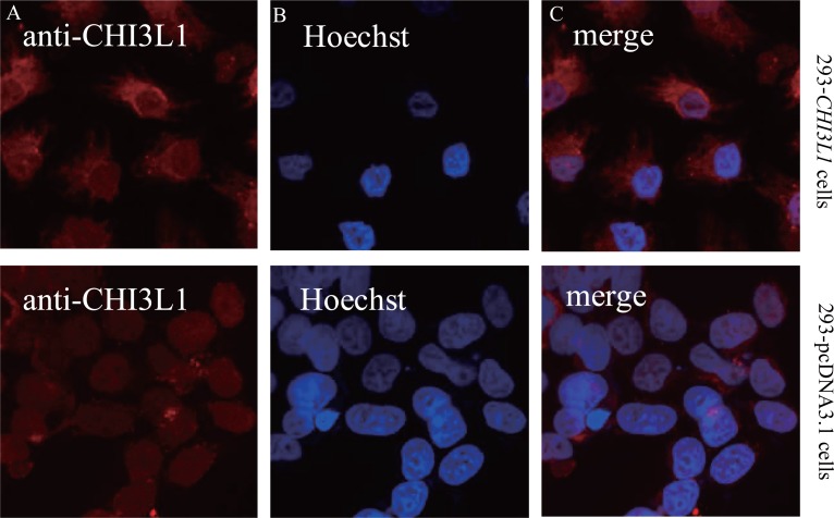
An important task in understanding oncogenesis is the identification of those genes whose copy number and expression increase during tumorigenesis. Previously, in an effort to identify genes which could be used as molecular markers for glial tumors, we compared gene expression in glioblastoma to the normal brain cells. Among the genes with the most pronounced increased expression in tumors there was CHI3L1, encoding the secreted chitinase 3-like 1 protein (also known as HC gp-39 or YKL-40). Expression of CHI3L1 was found increased significantly in various tumors in comparison with corresponding normal tissues. Here we show that CHI3L1 can decrease the doubling time of 293 cells. We have also demonstrated that CHI3L1 allows the anchorage-independent growth in soft agar and, in addition, stable CHI3L1 expression made 293 cells tumorigenic: these cells stimulate the initiation of tumors after their xenograft transplantation into the Wistar rat brains. Thus, the overexpression of CHI3L1 is likely to be critical in the development of some tumors and when we gain more information about mechanisms of CHI3L1 oncogenicity, it could be used as one of the potential targets for anticancer therapy.
Keywords: chitinase 3-like 1 protein (CHI3L1); glioblastoma; oncogene.
Figures


Similar articles
-
Chitinase-3 like-protein-1 function and its role in diseases.Signal Transduct Target Ther. 2020 Sep 14;5(1):201. doi: 10.1038/s41392-020-00303-7. Signal Transduct Target Ther. 2020. PMID: 32929074 Free PMC article. Review.
-
Two closely related human members of chitinase-like family, CHI3L1 and CHI3L2, activate ERK1/2 in 293 and U373 cells but have the different influence on cell proliferation.Int J Biol Sci. 2012;8(1):39-48. doi: 10.7150/ijbs.8.39. Epub 2011 Nov 16. Int J Biol Sci. 2012. PMID: 22211103 Free PMC article.
-
CHI3L1 (YKL-40) is expressed in human gliomas and regulates the invasion, growth and survival of glioma cells.Int J Cancer. 2011 Mar 15;128(6):1316-26. doi: 10.1002/ijc.25466. Int J Cancer. 2011. PMID: 20506295
-
The chitinase-like protein YKL-40 is secreted by airway epithelial cells at base line and in response to compressive mechanical stress.J Biol Chem. 2010 Sep 24;285(39):29817-25. doi: 10.1074/jbc.M110.103416. Epub 2010 Jul 22. J Biol Chem. 2010. PMID: 20650887 Free PMC article.
-
Uncovering novel mechanisms of chitinase-3-like protein 1 in driving inflammation-associated cancers.Cancer Cell Int. 2024 Jul 27;24(1):268. doi: 10.1186/s12935-024-03425-y. Cancer Cell Int. 2024. PMID: 39068486 Free PMC article. Review.
Cited by
-
Epithelial Membrane Protein-3 and Chitinase-3-like Protein-1 as New Prognostic Predictors of Glioma, a Two-Gene Study.Curr Oncol. 2023 Sep 23;30(10):8686-8702. doi: 10.3390/curroncol30100629. Curr Oncol. 2023. PMID: 37887529 Free PMC article.
-
Chitinase-3 like-protein-1 function and its role in diseases.Signal Transduct Target Ther. 2020 Sep 14;5(1):201. doi: 10.1038/s41392-020-00303-7. Signal Transduct Target Ther. 2020. PMID: 32929074 Free PMC article. Review.
-
Unveiling YKL-40, from Serum Marker to Target Therapy in Glioblastoma.Front Oncol. 2014 Apr 28;4:90. doi: 10.3389/fonc.2014.00090. eCollection 2014. Front Oncol. 2014. PMID: 24809021 Free PMC article. Review.
-
Dis3L2 regulates cell proliferation and tissue growth through a conserved mechanism.PLoS Genet. 2020 Dec 28;16(12):e1009297. doi: 10.1371/journal.pgen.1009297. eCollection 2020 Dec. PLoS Genet. 2020. PMID: 33370287 Free PMC article.
-
Two closely related human members of chitinase-like family, CHI3L1 and CHI3L2, activate ERK1/2 in 293 and U373 cells but have the different influence on cell proliferation.Int J Biol Sci. 2012;8(1):39-48. doi: 10.7150/ijbs.8.39. Epub 2011 Nov 16. Int J Biol Sci. 2012. PMID: 22211103 Free PMC article.
References
-
- Garifulin OM, Shostak KO, Dmitrenko VV, Rozumenko VD, et al. Increased expression of SOX-2 and HC gp-39 genes in astrocytic tumours. Biopol. Cell. 2002;18:324.
-
- Tanwar M, Gilbert MR, Holland EC. Gene expression microarray analysis reveals YKL-40 to be a potential serum marker for malignant character in human glioma. Cancer Res. 2002;62(15):4364. - PubMed
-
- Shostak K, Labunskyy V, Dmitrenko V, Malisheva T, et al. HC gp-39 gene is upregulated in glioblastomas. Cancer Lett. 2003;198(2):203. - PubMed
-
- Johansen JS. Studies on serum CHI3L1 as a biomarker in diseases with inflammation, tissue remodelling, fibroses and cancer. Dan. Med. Bull. 2006;53(15):194. - PubMed
-
- Riemenschneider MJ, Knobbe MJCB, Reifenberger G. Refined mapping of 1q32 amplicons in malignant gliomas confirms MDM4 as the main amplification target. Int. J. Cancer. 2003;104(6):752. - PubMed
LinkOut - more resources
Full Text Sources
Other Literature Sources
Research Materials