. 2012 Mar;8(1):51-63.

The Predominant Proteins that React to the MC-20 Estrogen Receptor Alpha Antibody Differ in Molecular Weight between the Mammary Gland and Uterus in the Mouse and Rat

Affiliations

The Predominant Proteins that React to the MC-20 Estrogen Receptor Alpha Antibody Differ in Molecular Weight between the Mammary Gland and Uterus in the Mouse and Rat

Aliccia Bollig-Fischer et al. Int J Biomed Sci. 2012 Mar.

Abstract

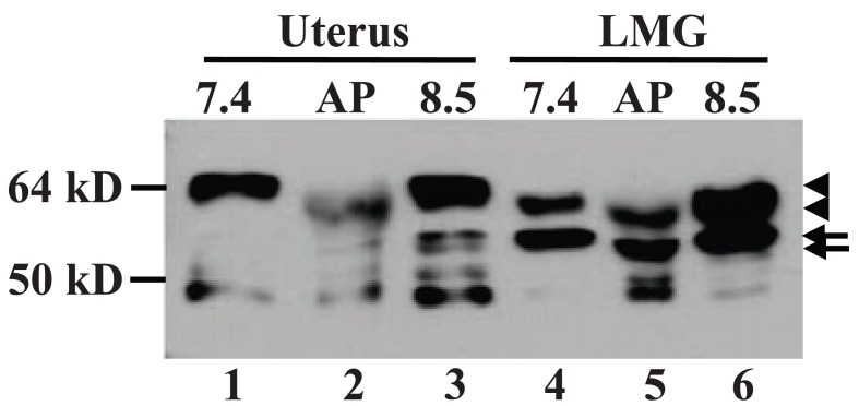
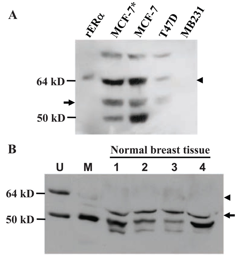
There are many estrogen receptor α (ERα) antibodies available but few of them target a rodent ERα. Using the MC-20 antibody raised against the C-terminus of mouse ERα, we show in this communication that in the mammary gland of female mice and rats, the wild type (wt) ERα was detected on immunoblots as a dominant protein only during lactation, and the protein was lactating specific as it migrated slightly faster than the 67-kD wt ERα in the uterus, likely due to a different phosphorylation status. In contrast, in the nulliparous, pregnant, involuting and involuted mammary glands, the dominant protein recognized by MC-20 was about 61-kD, which is dubbed herein as "MC-20 reactive protein" or MC20RP in abbreviation as its identity is unknown. Our results showed that it was not derived from proteolysis or de-phosphorylation of the 67-kD ERα and was unlikely to be translated from an ERα mRNA variant. Ovariectomy decreased the lactating specific wt ERα but increased the 61-kD MC20RP in the mammary tumors from MMTV-c-myc transgenic mice but these two proteins in the uterus were unaffected. The 61-kD MC20RP was decreased in the mammary tumors, compared with proliferating mammary glands, in estrogen-treated ACI rats. These results suggest that while the lactating specific wt ERα alone or together with the MC20RP may sustain lactation, the MC20RP may support proliferation of the mammary gland and some mammary tumors.

Keywords: breast cancer; estrogen receptor alpha; mammary gland; uterus.


Figures

Figure 1
Figure 1
Immunoblot detection of the 67-kD wild type (wt) ERα (arrowhead) and a 61-kD protein MC20RP (arrow). A: The 67-kD wt ERα is detected at high abundance in the uterus (U) by MC-20 antibody. However, the wt ERα in the mammary tissue (M) from a virgin CD rat and a FVB mouse is only faintly detected and, in the rat, it migrated slightly faster than its counterpart in the uterus (bottom-arrowhead in lane 2 vs top-arrowhead in lane 1). In the mammary tissue, the dominant protein recognized by MC-20 is estimated as 61-kD, although it also migrates slightly slower in the mouse than in the rat (top-arrow in lane 4 vs bottom-arrow in lane 2). The second band in the mouse uterus might be the uterine 61-kD MC20RP, but it cannot be sure as it migrates slightly faster than the 61-kD MC20RP in the mouse mammary tissue. The band at about 50-kD in the mouse and rat uteri is an ERα isoform that has been well described in the literature and is not the focus herein (5, 10, 22-24). B: The 67-kD wt ERα was detected by MC20 only in the mouse uterus (U), not in the mammary tissue from five virgin female FVB mice (M1-M5). In the mammary tissue, the protein detected is smaller than that in the uterus and its abundance varies greatly among the five animals, but comparison among samples should be made with caution because virgin mammary tissue is dominated by fat-tissue and varies greatly in the amounts of other cell types. C: The Ab-17 antibody raised against the N-terminus of human ERα can recognize the wt ERα in mouse uterus (U), not the 61-kD protein in the mouse mammary tissue (M). The blot is relatively murky because a slightly excessive amount of antibody was used to ensure that its affinity to the 61-kD MC20RP is indeed poor. D: Comparison of the sequences of the last 20 amino acids of the mouse, rat and human ERα. These 20 amino acids in the mouse and rat are identical and are used as the immunogen for the MC-20 antibody, whereas the last 15 amino acids are used as the immunogen for the C1355 antibody. The human sequence used as the immunogen of the HC-20 antibody differs from that of the mouse and rat by six amino acids that are boldfaced and underlined. Figures 1A and 1C are representative of similar analyses of at least three independent samples.
Figure 2
Figure 2
Immunoblot detection, with MC-20 antibody, of ERα in different developmental stages of the mammary gland and uterus of FVB mice. A and B: Pregnant (P) or lactating (L) mammary tissue and uterine tissue (U) were homogenized independently and in combination (U+P and U+L) at a 1:1 ratio. The uterine-derived 67-kD ERα (top-arrowhead) remains intact while the 61-kD MC20RP (arrow) remains unchanged in the U+P or U+L, relative to the level in the P or L alone. The weakly expressed wt ERα in pregnant mammary glands (bottom-arrowhead in lane 2 in A) migrates slightly faster than the wt ERα (top-arrowhead in lane 1 in A) the uterus. C: The 67-kD ERα (arrowhead) and the 61-kD MC20RP (arrow) in the uterus as well as in the nulliparous, involuting (2-day post-weaning) and involuted (two-month post-weaning) mouse mammary gland. D: Detection of the 67-kD ERα (arrowhead) and the 61-kD MC20RP (arrow) in mammary glands and uteri from virgin (V), lactating (L) or pregnant (P) mice. Figures 2A and 2B are representative of analyses of three independent samples for each stage of development. Figure 2C and 2D are representative of analyses of two independently collected tissue sets.
Figure 3
Figure 3
Phosphorylation status of ERα proteins. Immunoblot analysis of proteins extracted from uterus and lactating mammary glands (LMG) with lysis buffer at pH7.4 or 8.5. A portion of pH8.5 extract was incubated with calf intestine alkaline phosphatase (AP) for 1 h followed by SDS-PAGE immunoblot analysis. The higher pH condition does not change the 67-kD ERα migration (top-arrowhead in lanes 1 vs 3 and lanes 4 vs 6), but the 67-kD ERα in the AP-treated extracts from both organs is down-shifted compared with its counterpart in the non-treated extract at pH8.5 (bottom-arrowhead in lanes 2 and 5 vs top-arrowhead in lanes 3 and 6, respectively). Note that the AP-treated wt ERα in the uterus is down-shifted to the position of the non-treated wt (lactating specific) ERα in the LMG (bottom-arrowhead in lanes 2 vs 4 and 6). AP treatment also down-shifts the 61-kD MC20RP in the LMG (top-arrow in lane 6 vs bottom-arrow in lane 5). The experiment was run three times.
Figure 4
Figure 4
The ratio of the 67-kD wt ERα to the 61-kD MC20RP. A: The ratio of the 67-kD ERα (bottom-arrowhead) to the 61-kD MC20RP (arrow) is about 1.5:1 in lactating mammary glands (LM) but is about 1:2 in an MMTV-c-myc mammary tumor (MT). A uterus (U) included as a reference shows the abundantly expressed wt ERα, which is at a slightly higher molecular position than the wt ERα in the LM and MT (top-arrowhead vs bottom-arrowhead). B: When tumor bearing MMTV-c-myc mice were ovariectomized two weeks prior to the tumor collection (ovx tumors), the 67-kD wt ERα (arrowhead) level is decreased while the 61-kD MC20RP (arrow) level is increased, leading to a dramatic decrease in the ratio of the 67-kD to 61-kD proteins, compared with the ratio seen in intact animals shown in panel A. The ratio is unaffected in the uteri of ovariectomized non-transgenic mice, compared with the intact mice (lanes 1 vs 2), while nulliparous mammary tissue from the same ovariectomized mice shows only the 61-kD MC20RP. The figure represents analyses of tumors from three intact mice and two ovariectomized tumor bearing mice and two non-transgenic mice.
Figure 5
Figure 5
The effect of estrogen-enhanced ubiquitin-proteosome pathway on the 67-kD ERα and the 61-kD MC20RP. A: myc-MT1 cells were cultured in DMEM containing 5% charcoal-treated calf serum for 1 week prior to assay. After 1 h pre-treatment with 1 mM MG132, a proteosome inhibitor, or with DMSO vehicle, cells were treated for 16 h with 5 nM 17β-estradiol (E2), 5 μM tamoxifen (Tam), or ethanol (vehicle) as indicated. Cell lysates were then prepared and analyzed by immunoblot. The 61-kD MC20RP (arrow) is the only protein detected when the film was exposed for an appropriate time (bottom panel). The 67-kD ERα (arrowhead) is detected only when the film is overexposed (top panel). Note that while the 61-kD MC20RP is unaffected by MG132, E2 or tamoxifen, the 67-kD ERα is decreased by E2 in the absence of MG132 (lane 3 in the top panel). The experiment was performed twice. B: DES-induced proliferating mammary tissue (DES) or mammary tumors (MT) from female ACI rats and normal mammary tissue (Con) from non-treated rats do not express detectable amount of the 67-kD ERα (arrowhead) that is abundantly expressed in the uterus (U) from a non-treated ACI rat included as a positive control. The 61-kD MC20RP (arrow) is expressed in non-treated and DES-treated mammary tissue but is barely detectable in the tumors. Comparisons of the expression levels among different samples should be made with caution because of the great difference in the cellularity, i.e. the contents of epithelial and adipose cells, although some ERα protein isoforms with lower molecular weights may be used as internal reference.
Figure 6
Figure 6
Immunoblot detection of the 61-kD MC20RP in a panel of mouse organs. These samples were resolved by a less rigorous electrophoresis (on an 8 cm, 12% acrylamide gel) than those in previous figures. The degree of separation between the 67-kD wt ERα (arrowhead; the uppermost band) and the MC20RP just beneath it (arrow) is narrowed with this more routine approach of SDS-PAGE. Note that the MC20RP is expressed in all of these tissues but the 67-kD ERα is mainly detected in the uterus, brain, kidney and mammary tissue (Mam). The figure represents analyses of two independently collected sample sets.
Figure 7
Figure 7
Detection of human MC20RP analog. A: Western blot assay with HC-20 ERα antibody and total lysates (100 μg proteins) from ERα positive MCF7 and T47D and negative MDA-MB231 (MB231) human breast cancer cell lines. MCF-7 cells were cultured with additional 4 μg/ml insulin or without (MCF-7*) it, as insulin may affect MCF-7 cell growth and differentiation. Purified human recombinant ERα (rERα) protein is included as a control due to the unavailability of endometrium-enriching human uterus sample. The arrow indicates a band migrating similarly to the 61-kD MC20RP, whereas arrowhead indicates the wt ERα. B: Western blot assay with HC-20 antibody and 100 μg protein lysates of uninvolved (i.e. relatively normal) breast tissue from four breast cancer patients. Uterine (U) and mammary (M) tissues from a virgin mouse are included as controls. Note that the uninvolved breast tissues show a band migrating similarly to the 61-kD MC20RP (arrow). The HC-20 antibody can detect readily the 67-kD wt ERα (arrowhead) in mouse uterus but it detects only faintly the 61-kD MC20RP and also the wt ERα in the mouse mammary tissue.

Similar articles

Cited by

References

    1. Stingl J. Estrogen and progesterone in normal mammary gland development and in cancer. Horm. Cancer. 2011;2:85–90. - PMC - PubMed
    1. Russo J, Russo IH. The role of estrogen in the initiation of breast cancer. J. Steroid Biochem. Mol. Biol. 2006;102:89–96. - PMC - PubMed
    1. Nicholson TM, Ricke WA. Androgens and estrogens in benign prostatic hyperplasia: past, present and future. Differentiation. 2011;82:184–199. - PMC - PubMed
    1. Thomas C, Gustafsson JA. The different roles of ER subtypes in cancer biology and therapy. Nat. Rev. Cancer. 2011;11:597–608. - PubMed
    1. Liao DZ, Pantazis CG, Hou X, Li SA. Promotion of estrogen-induced mammary gland carcinogenesis by androgen in the male Noble rat: probable mediation by steroid receptors. Carcinogenesis. 1998;19:2173–2180. - PubMed

LinkOut - more resources